Download
1 / 50
500 likes | 619 Views
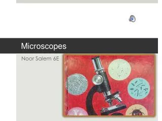
MICROSCOPES. Microscope Quiz Friday –Jan. 28. Label parts of microscope How to use (ex: use coarse knob to find object, adjust diaphragm for light) Total Magnification (eyepiece X objective) How to Measure ( field diam. number across)

E N D
Microscope Quiz Friday –Jan. 28 • Label parts of microscope • How to use (ex: use coarse knob to find object, adjust diaphragm for light) • Total Magnification (eyepiece X objective) • How to Measure (field diam. • number across) • Convert mm to µm (4 mm = 4000 µm)
Compound Light • Uses two lenses • ocular • objective • To bend light
Resolving Power • Being able to tell two objects apart • Measure of “clarity”-how clear it is
Resolving Power • smallest separation between two object points that a given lens (or mirror) can still show as two distinct entities, not one • .. .
Pollen Under 1000X LM Over 1000 X SEM
MAGNIFICATION • Increase in the apparent size of an object • MULTIPLY THE OCULAR LENS x THE OBJECTIVE • OCULAR 10x • OBJECTIVE 40 x • WHAT IS THE TOTAL MAGNIFICATION? • 400 x
Leaf 4X Leaf 10 X How do they look different?
ADVANTAGES of CLM: • CAN MAGNIFY UP TO 1000 x • CAN VIEW LIVING THINGS • Resolving power 200 nm or 0.2 µm
Disadvantages of LM • Objects must be thin or transparent so light can go through them • The image is inverted
Pictures of LM microorganisms Can be stained
Dissecting Light Microscope • Image is NOT inverted • Usually 40 X is the limit
Dissecting Scope Viewing Light-colored stage for dark specimens and dark-colored stage for light ones
ELECTRON MICROSCOPES • USE MAGNETS TO FOCUS A BEAM OF ELECTRONS • TEM (Transmission Electron Microscope) • SEM (Scanning Electron Microscope)
TEM AdvantageCan magnify 1000 X’s more than a light microscope (Uh…1000 X 1000 = 1,000,000 X’s ) • Resolving power 0.2 nm
TEM Disadvantages • Must be in a vacuum (dead) • Sample must be VERY THIN (less than 0.2 nm)
SEM: Scanning ElectronMicroscope Advantages • Electron beam scans the surface • Resolution 10 nm • Magnifies 1,000,000 X’s
SEM Disadvantages • Must be in a vacuum (dead) • Cannot see internal structures
SEM Images • house fly • and its mouth
Choose Critter and Change Image • Molecular Expressions Microscopy Primer: Electron Microscopy Interactive Java Tutorials - Virtual Scanning Electron Microscopy
SPM-Scanning Probe • Scanning Probe Microscope • Viewed ATOMS!!!!!!!!!!! • Does not need sample in a vacuum • Magnifies 10 million times
SPM Images (50 um X 1.4 um) • Steel Surface
SPM Images • DNA
CHOICES: Diaphragm Objectives Ocular Coarse knob Fine knob Base Revolving nose piece Stage and stage clips Arm condenser Label the parts:
Label the parts: Ocular (eyepiece) Arm Coarse knob Fine knob (little) Revolving nose piece Objectives Stage and stage clips Diaphragm Condenser base
That is, as you increase magnification, the actual field of view becomes proportionally smaller. • 4OX 100X 400X
Viewing “F” with A Light Microscope • Which is an “F” put in a compound light and a dissection light microscope?
Field of View • What is the approximate width in mm? • In µm? (1 mm = 1000 µm) • 4 mm 4000 µm
Do the math: • one millimeter (mm.) = 1,000 micrometersµm • So 5.5 mm = ________ µm • 5500 µm
NOTE!!!!!!!!!!!!!!!! • The field diameter at high power is proportional to the ratio of the low to high power objectives. • If 40X is 4000 µm • 400X is 400 µm
FD = field diameter • Low power FD X low magnification high power magnification = high power FD Use when object is between the mm markers
Why do you need to know Field Diameter? • You may wish to estimate the size of the specimens (e.g., cells) you will see in lab.
Field of View • What is the approximate size of this cell? • In mm? • In um? • If 5 fit across… • O.4 mm 400 µm
If the field of view in this question is 2 mm… How long is one cell? 2 mm
If the field of view in this question is 2 mm… If 3 cells fit across then, one cell is: 2mm 3 =.67 mm 2 mm
Wet Mount 1. Add drop of water 3. Add cover slip 2. Place Specimen on 4. Tap out bubbles slide
Making a Wet Mount Slide • Add drop of water • Add cover slip