Download
1 / 13
130 likes | 155 Views
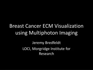
Learn how to visualize breast cancer extracellular matrix using advanced multiphoton imaging techniques. Follow the detailed protocol for staining and imaging samples to study different tissue structures and cancer types.

E N D
Breast Cancer ECM Visualization using Multiphoton Imaging Jeremy Bredfeldt LOCI, Morgridge Institute for Research
H&E and SHG • Protocol • Thaw & fix sample • Section sample to 200 microns on a vibratome • Image with SHG • Paraffin embed tissue wafer • Section and stain for H&E • Slide scan • Green = SHG signal (Collagen Type I)
Normal Associated Tissue H&E SHG 11.6 mm
Invasive Ductal Carcinoma SHG H&E
Fibroadenoma H&E SHG
Mucinous Carcinoma 7.5 mm H&E SHG
Intrinsic Fluorescence • Green = SHG • Red = Autofluorescence from FAD or Collagen type III
Extrinsic Fluorescence • Green = SHG (Collagen type I) • Red = E-Cadherin stained cells & Autofluorescence from Collagen type III
Terminal Ductal Lobular Unit (Red) Surrounded by Collagen (Green)
IDC Cells Near a Vessel with ECM IDC Cells Vessel