Understanding Cells: The Basic Units of Life and Their Complexities
Cells are the fundamental units of life, responsible for various functions including metabolism, growth, repair, and reproduction. According to Cell Theory, proposed by scientists like Schleiden, Schwann, and later expanded by Virchow, all living organisms are composed of cells. This overview explores the general characteristics of cells, differences between prokaryotic and eukaryotic cells, and how cells can be studied using various types of microscopes and techniques. We also discuss exceptions to the cell theory, including the unique features of mitochondria, chloroplasts, and viruses.


Understanding Cells: The Basic Units of Life and Their Complexities
E N D
Presentation Transcript
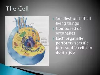
I. Definition: The smallest (basic) unit of life. It carries out: A. metabolism B. Growth C. Repair D. reproduction
II. Cell Theory: Matthias Scleiden and Theodore Schwann A. The Cell: 1. Is the basic unit of life. 2. Is the basic unit of function. 3. 1855 – expanded by Virchow – all cells come frompreexisting cells – He observed mitosis.
III. Exceptions to the Cell Theory: A. Mitochondria B. Chloroplasts C. Multinucleated cells D. Prokaryotes E. Viruses
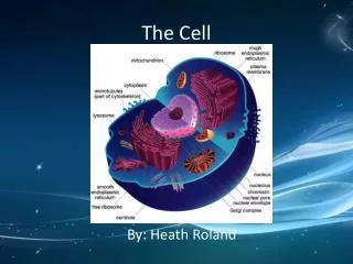
IV. General Characteristics: A. Nuclear material or DNA B. Cytoplasm (can have organelles) C. Cell membrane
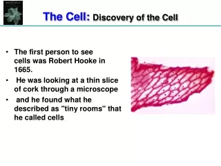
V. How Cells are Studied: A. History: 1. 1632 – 1723 Anton Van Leeuenhoek a. microscope maker b. observed “wee little beasties” in pond H2O (observed living material) 2. 1665 – Hooke a. coined term cell b. observed non-living cork cells 3. By 1800’s Scientists founded and described manyorganelles.
B. Microscopes: 1. Simple hand lens – mag. 10-15x 2. Compound – 2 sets of lenses a. ocular lens = 10x b. objectives = 4x, 10x, 40x, 100x c. One observes the gross anatomical characteristics of cells and tissues.
3. Electron Microscope – studies cells only. T.E.M. = Transmission Electron Microscope a. uses magnetic lenses instead of glass b. uses electron beam instead of white light c. mag. 250,000x or more d. use: internal cell characteristics S.E.M. = Scanning Electron Microscope a. uses 3D – see morphology of cells – outsidecharacteristics (shape)
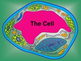
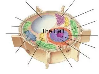
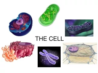
Plant Cell:
C. Additional Studies: 1. Physically – centrifugation 2. biochemically – chromatography, electrophoresis
VI. Basic Cell Types: A. Prokaryotic cell: 1. representative of K. Monera, D. Archaea, Bacteria 2. has a nucleoid area (DNA is naked) no protein associated with it. 3. lacks other membranous organelles 4. plasma membrane is the site of: a. cell respiration b. cellular photosynthesis 5. has Ribosomes – smaller than Eukaryotes: a. are the sites of protein synthesis
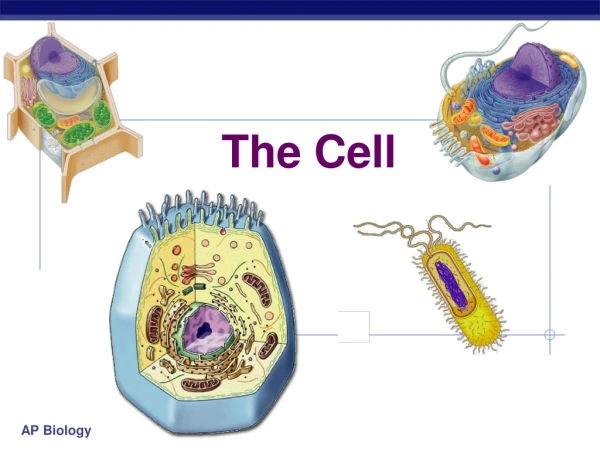
B. Eukaryotic Cells (true nucleus): 1. typical of 4 other kingdoms 2. distinct nucleus surrounded by nuclear membrane 3. membrane bounded organelles Difference between Plant and Animal Cells:
Plant Animal 1. Cell Wall none 2. Plastids none a. chloroplast b. chromoplast c. leucoplasts 3. large central vacuole none 4. none centrioles 5. none found lysosomes
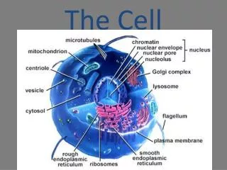
VII. Internal Cell Characteristics: A. Non-living Parts (outline) 1. cell walls: a. Monera b. Protista c. fungi d. plantae 2. middle lamella, plant cells
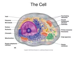
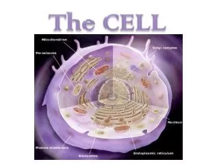
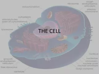
B. Living Parts = Protoplasm 1. Cell membrane (Plasma) 2. Cytoplasm 3. Endoplasmic Reticulum 4. Ribosome 5. Golgi Bodies 6. Lysosomes 7. Plastids
8. Microbodies (peroxisomes) 9. Mitochondria 10. Cytoskeleton 11. Centrosome – Centrioles -Basal Bodies 12. Cilia and Flagella 13. Vacuoles and Vesicles 14. Nucleus incl. Nucleolus
C. Non-Living Parts: 1. Cell Walls: a. found in 4 out of 5 kingdoms b. Monera: 1. Bacteria – cell walls, protein and fats 2. Blue-green algae, cell walls – cellulose c. Protista: 1. Algae – cell wall mostly cellulose d. Fungi: cell wall are cellulose-chitinous e. Plantae: cell wall are cellulose
Remember: f. Cellulose = complex polysaccharide g. In Plantae – cell walls can be thick and several layers (Secondary walls) 2. Middle lamella: a. cements plant cells together b. composed of mostly calcium pectate and other polysaccharides.
D. Living Parts = Protoplasm (Elaborated) 1. Cell membrane: a. is fluid b. composed of a: 1. phospholipid bilayer 2. proteins – embedded in membrane = mosaic pattern c. is differentially permeable
d. its function: 1. to bind and transport materials into andout of the cell. 2. communicate with other cells. 3. chemicals can attach to it causing a r’xn or change (ex: hormonal attachment) 4. gives cell it shape
2. Cytoplasm: a. semi fluid composed of 70-90% water b. is the site of cellular work: 1. such as cytoplasmic streaming, energy transfer reactions 2. contains organelles, some being anchored by filaments and move about on a Cytoskeleton (will treat later on) 3. the nucleus remains fixed
Endomembrane System • Endoplasmic reticulum • Golgi • Vacuoles—will be treated later • Plasma membrane--covered
3. Endoplasmic Reticulum: a. it is maize of internal tubular membranes b. it connects the nuclear membrane with the plasma membrane c. it divides the cytoplasm into compartments d. provides an increased surface area for enzymes and chemical r’xns to function.
e. Functions: 1. framework for r’xns 2. transport materials within the cell and from one cell to another 3. provides a temporary storage area for proteins 4. compounds are synthesized here.
f. Two types of E.R: 1. Smooth E.R.: a. produces lipids ex. Oils, phospholipids, steroids (sex hormones and adrenal steroids b. produces cellular secretions c. involved in carbohydrate metabolism glycogen (liver glucose phosphate) removes the phosphate glucose can leave the cell d. functions in the detoxification of drugs (ex. Liver) alcohols and barbiturates e. Involved in muscle contraction and the movement of Ca++ from ER to cytoplasm to trigger a contraction
2. Rough E.R.: abundant in cells that secrete proteins a. associated with ribosomes (bounded ribosomes) b. transports the proteins that are produced on their assoc.ribosomes. c. proteins can form their final shape (structures) here and/or can go to perhaps the Golgi to be finished. d. generic glycoproteins are produced here *oligosaccharides are added to the protein e. RE is abundant in secretory cells f. Membrane proteins are synthesized here as well as phospholipids (it is a membrane factory)
4. Ribosomes: a. composed of RNA and protein b. some are attached to E.R. and some are free in the cytoplasm c. are found in both prokaryotes and Eukaryotes d. function – provides the work bench for protein manufacturing e. can be in groups of 5 or more strung together = polysomes.
5. Golgi Bodies: (Complex) a. Packaging Plants b. Stacks of Plate like membranes and vesicles Filled with cell products. c. In animal cells they are located at one side of the nucleus. They are abundant in secretory cells d. trans – shipping: see above figure. And cis-receiving side e. Example-Protein (ER) -> sealed in a vesicle Enters into Golgi -> membrane fusion -> Protein may get a rearrangement of carbohydrate group (modified) -> stored -> protein secretion is concentrated -> released in a vesicle -> plasma memb. (Tags, sorts, sends)
Summary of Functions of the Golgi Body: Secretory cells – concentrates substances tobe secreted and delivers it to its destination outside of the membrane. They engage in synthesis all along the way: a. polysaccharides: maybe synthesized from simple sugars b. glycoproteins: are finished here c. proteins maybe stored modified andreleased into the cytoplasm. d. glycolipids: can be formed from phospholipids
Non-Secretory cells – Packages intracellular digestive enzymes into little organelles called the lysosomes Lysosomes are actually finished here but begin inthe ER 6. Lysosomes – Suicide sacs of cell pH 5 – work the best. a. covered with a single membrane b. contain hydrolytic enzymes for digestion. 1. scavengers – digest worn out parts or cells (phagocytosis) 2. Autodigestion burst – release digestive enzymes and can digest the cells they’re in (self destructive mechanism)
EX: Tad pole – absorbs its tail “self- destructive system following cellular death.” EX: Phagocytes – digest invading bacteria.