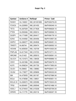
Suppl. Fig. 2
Suppl. Fig. 2. A. Cntrl. FasL 25ng/ml. B. 0.5 FasL +zVAD. 25 FasL + α -Fas. 25 FasL+ zVAD. 0.5 FasL +zVAD. 25 FasL+ zVAD. 25 FasL +zVAD. 0.5 FasL. 25 FasL. 0.5 FasL. 25 FasL. 5 FasL. α -Fas. zVAD. HuT78. Cntrl. Cntrl. zVAD. 0.5 FasL. Cntrl. 25 FasL. zVAD. Cntrl.

Suppl. Fig. 2
E N D
Presentation Transcript
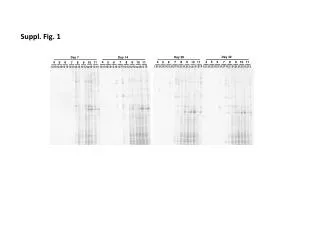
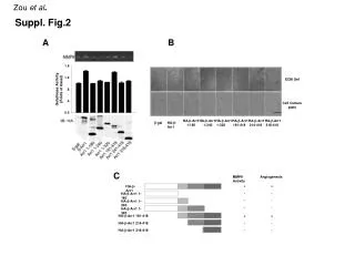
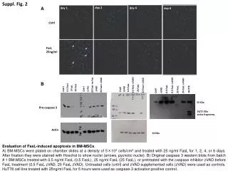
Suppl. Fig. 2 A Cntrl FasL 25ng/ml B 0.5 FasL +zVAD 25 FasL +α-Fas 25 FasL+ zVAD 0.5 FasL +zVAD 25 FasL+ zVAD 25 FasL +zVAD 0.5 FasL 25 FasL 0.5 FasL 25 FasL 5 FasL α-Fas zVAD HuT78 Cntrl Cntrl zVAD 0.5 FasL Cntrl 25 FasL zVAD Cntrl 35 KDa Pro-caspase 3 19/17 KDa active fragments Actin 43 KDa Evaluation of FasL-induced apoptosis in BM-MSCs. A) BM-MSCs were plated on chamber slides at a density of 5×10³ cells/cm2 and treated with 25 ng/ml FasL for 1, 2, 4, or 6 days. After fixation they were stained with Hoechst to show nuclei (arrows, pycnotic nuclei). B) Original caspase 3 western blots from batch # 1 BM-MSCs treated with 0.5 ng/ml FasL (0.5 FasL), 25 ng/ml FasL (25 FasL), or pretreated with the caspase inhibitor zVAD before FasL treatment (0.5 FasL zVAD, 25 FasL zVAD). Untreated cells (cntrl) and zVAD-supplemented cells (zVAD) were used as controls. HuT78 cell line treated with 25ng/ml FasL for 5 hours were used as caspase-3 activation positive control. day 6 day 2 day 1 day 4 day 6