Download
1 / 29
300 likes | 785 Views
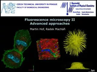
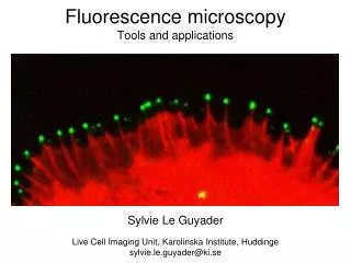
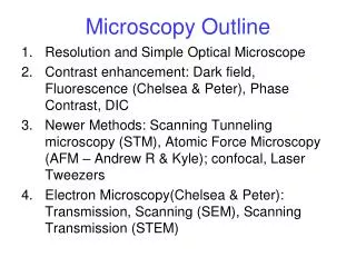
Fluorescence Microscopy: Membranes and Pharmaceuticals. Nicole Antczak University of Connecticut CHEM 5395 March 3, 2001. Outline. Fluorescence Overview Fluorescence Microscope Confocal Micoscopy Fluorophores FRET Torchilin study. Fluorescence – Jablonski Diagram.

E N D
Fluorescence Microscopy: Membranes and Pharmaceuticals Nicole Antczak University of Connecticut CHEM 5395 March 3, 2001
Outline • Fluorescence Overview • Fluorescence Microscope • Confocal Micoscopy • Fluorophores • FRET • Torchilin study
Fluorescence – Jablonski Diagram http://www.olympusmicro.com/primer/java/jablonski/jabintro/index.html
http://www.olympusmicro.com/primer/techniques/fluorescence/anatomy/bx51fluorescence.htmlhttp://www.olympusmicro.com/primer/techniques/fluorescence/anatomy/bx51fluorescence.html
http://www.olympusmicro.com/primer/techniques/fluorescence/anatomy/ix70fluorescence.htmlhttp://www.olympusmicro.com/primer/techniques/fluorescence/anatomy/ix70fluorescence.html
Confocal Microscopy http://www.olympusmicro.com/primer/techniques/confocal/confocalintro.html
Confocal Microscopy1 • Uses laser scanning • Limited number of laser wavelengths • Allows for scanning not only in x-y but in z direction • produce thin (0.5 to 1.5 micrometer) optical sections through fluorescent specimens that have a thickness ranging up to 50 micrometers or more
http://www.olympusmicro.com/primer/techniques/confocal/confocalintro.htmlhttp://www.olympusmicro.com/primer/techniques/confocal/confocalintro.html
With Software… http://www.olympusmicro.com/primer/techniques/confocal/confocalintro.html
http://www.olympusmicro.com/moviegallery/confocal/rk13mkoh2b/index.htmlhttp://www.olympusmicro.com/moviegallery/confocal/rk13mkoh2b/index.html
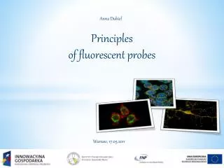
Fluophores • Fluorescein/DAPI/Texas Red • GFP and related proteins • Quantum Dots
Fluorescence Resonance Energy Transfer • Occurs between donor molecule and acceptor molecule • Can be used to measure distance between two sites on a macromolecule • Also used to detect protein-protein interactions • Membrane fusion and lipid exchange
Torchilin Study5 Fluorescence microscopy to follow the targeting of liposomes and micelles to cells and their intracellular fate
Liposomes5 • Artificial phospholipid vesicles • Loaded with water soluble drugs or water insoluble drugs • Typically use antibodies for targeting • Long circulated liposomes coated with PEG • Why not combine the two?
Micelles5 • Colloidal dispersions of particle size within to 5 to 100nm • Able to increase solubility and bioavailability of poorly soluble pharmaceuticals • Several key micelle properties for drug incorporation • Size, CMC, load capacity of hydrophobic core • Small size advantageous • PEG/antibodies
Enhancing Micelle Cellular Uptake • Cationic lipid formulations Lipofectin (LL) • PEG-PE micelles carry negative charge • Combination of both could improve uptake
Delivery of Vitamin K3 Micelles • Vitamin K3 (VK3) inhibits growth of various cancer cell types • May be involved in arylation of cellular thiols • DBU used to accelerate formation of thioether of VK3 analogs • Does it have synergystic effects? • Prepared micelles with both DBU and VK3 and micelles with just VK3
TAT-Liposomes • Trans-activating transcriptional activator protein (TAT) • Attached TAT-peptide to liposomes via PEG/PE molecule • Several hundred TAT-peptides with single 200nm liposome
Testing integrity of TAT-Lipsome Complex • Rh-PE labeled liposomes with entrapped FTIC-dextran • Colocalization of Rh and FTIC fluoresence
Conclusions • Can be used to follow targeting of liposomes and micelles • Can be used for live cell imaging
Citations • http://www.olympusmicro.com/index.html • http://www.olympusmicro.com/primer/techniques/fluorescence/fluorhome.html • http://www.olympusmicro.com/primer/techniques/confocal/index.html • Lakowicz • Torchilin, V. P. Advanced Drug Delivery Reviews 57 (2005) 95– 109