Download
1 / 56
560 likes | 601 Views
Explore the intricate details of the central and peripheral nervous system, including the brain, spinal cord, and nerve pathways. Learn about the functional divisions, meninges, spinal cord segments, and more.

E N D
Nervoussystem: brain, spinalcord Dr. L. Kiss Anna Department of Anatomy, Histology and DevelopmentalBiology, Semmelweis University 2019
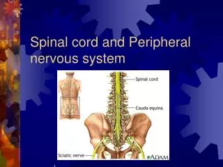
Nervous system Centralnervoussystem Peripheralnervoussystem Spinalcordbrain Spinalnerves, cranialnerves, ganglions • Functionally: somatic (axialmuscles) • vegetatíve: (internalorgans)
Reflex arch center afferens efferens efferens afferens receptor effector
Functional division of the nervous system Somatic Autonomic (vegetative) • voluntary - unvoluntary • starts and stops functions - regulates the functions (slow down-speed up) • segmental - network sympatheticparasympathetic thora-columbar cranio-sacral
Spinal cord: meningeal layers • Dura mater: outerlayer: endorachis innerlayer • Arachnoidlayer • Pia mater Epidural spce Subdural space Subarachnoid space
MENINGES Dura mater Arachnoid Pia mater: Denticulate ligament
Meningeallayers of thespinalcord ganglion spinale arachnoidea filaradicularia dorsalia dura mater ligamentum denticulatum
MENINGES Epidural space Subarachnoid space Denticulate ligament
MACROSCOPY Enlargements: Cervical enlargement (C4-T1) Lumbosacral enlargement (L2-S3) Conus terminalis Cauda equina Filum terminale Central nervous system Brain Spinal cord Peripheral nervous system Nerves Ganglia
Bloddsupply of thespinalcord radicular arteries vertebral artery subclavian artery radicular arteries Aortic arch pathway of the vertebral artery
Vasocoronamedullaris • Vertebralartery: • a.spinalisanterior (united) • a spinalisposterior (2) • Radiculararteries: • cervical: vertebral a. • ascending. cerv. a. • deepcerv. a. • thoracal: intercostalaa. • lumbal: lumbalaa. • sacral: lateralsacr. art.
CROSS SECTION Dorsal median sulcus Gray matter White matter Ventral median fissure Central canal
Crosssection of thespinalcord post. (dors.)median sulcus post. (dorsalis)horn intermediatezone (Th and L2-L3): lat.horn centralcanal ant. (ventral) horn ant. (ventr.) medianfissure
Spinal cord Dorsalcollumn (posterior): mainlysensorypathways Ventralcollumn (anterior): mainly motor pathways Lateralcollumn: mainlyvegetativpathways
SPINAL NERVES Dorsal branch Ventral branch
SPINAL CORD SEGMENTS Spinal cord ends at the level of the L1 vertebra in adults Lumbar and sacral roots travel several vertebrae down: cauda equina
SPINAL CORD SEGMENTS Number of segments: 8 cervical (!) 12 thoracic 5 lumbar 5 sacral 1 coccygeal
SEGMENTAL INNERVATION Dermatomes: segmental symptomes
CERVICAL PLEXUS AND BRACHIAL PLEXUS Cervical plexus (C1-4): Phrenic nerve Branchial plexus (C5-T1): Superior trunk (C5-6) Middle trunk (C7) Inferior trunk (C8-T1)
CERVICAL PLEXUS AND BRACHIAL PLEXUS Branchial plexus (C5-T1): Lateral cord (C5-7) Medial cord (C8-T1) Posterior cord (C5-T1)
LUMBOSACRAL PLEXUS Lumbar plexus (T12-L4): Sacral plexus (L4-S3):
Brain Medulla oblongata Pons Midbrain (mesencephalon) Cerebellum Diencephalon (thalamus, hypothalamus, hypophysis) Cerebrum Brain stem
MEDIANSAGITTAL SECTION OF THE BRAIN STEM M M Mo P Mo
Meningeal layers of the brain Dura mater: falx cerebri, tentorium cerebelli, falx cerebelli sinuses: venous blood Arachnoid layer: cisterns CSF Pia mater: blood vessels
VENTRICLES Lateral ventricle 4th ventricle 3rd ventricle
CEREBROSPINAL FLUID Production: choroid plexus: into ventricles Absorption: arachnoid villi: from subarachnoid space into superior sagittal sinus
BRAIN: Cerebrum SUPERIOR VIEW Frontal lobe Longitudinal cerebral fissure Parietal lobe Occipital lobe
BRAIN: LATERAL VIEW Parietal lobe Central sulcus Frontal lobe Occipital lobe Transverse cerebral fissure Lateral cerebral sulcus Cerebellum Temporal lobe Pons Medulla oblongata
BRAIN: MEDIANSAGITTAL CUT Longitudinal cerebral fissure Parietal lobe Corpus callosum Occipital lobe Frontal lobe Transverse cerebral fissure Cerebellum Temporal lobe
praecentral gyrus central sulcus postcentral gyrus
BRAIN: INFERIOR VIEW Longitudinal cerebral fissure Olfactory nerve (I) Frontal lobe Optic nerve (II) Optic chiasm Temporal lobe Optic tract Diencephalon Pons Medulla oblongata Cerebellum Occipital lobe
BLOOD SUPPLY: ARTERIES Arterial supply: Vertebral artery Internal carotid artery internal carotid artery vertebral artery
CIRCLE OF WILLISI Anterior communicating a. Anterior cerebral a. Internal carotid a. Middle cerebral a. Posterior communicating a. Posterior cerebral a. Basilar a. Vertebral a.
BLOOD SUPPLY: VEINS Venous drainage: Internal jugular vein: Sinuses Cerebral veins
BRAIN STEM: MEDULLA Glossopharyngeal n. (IX) Vagus n. (X) Olive Accessory n. (XI) Pyramid Hypoglossal n. (XII)
BRAIN STEM Cerebral aqueduct Cerebellum Midbrain Pons Fourth ventricle Medulla Central canal
CEREBELLUM Cerebellar hemisphere Vermis Tonsil
CEREBELLUM Fastigial nucleus Cerebellar cortex Globose nucleus Emboliform nucleus White matter Dentate nucleus