Download
1 / 32
390 likes | 1.41k Views
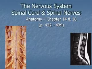
The Nervous System Spinal Cord & Spinal Nerves. Anatomy – Chapter 14 & 16 (p. 432 - 439). The Central Nervous System. The CNS is well protected by bone, CT, and fluid. Meninges – Connective tissues that surround and protect the brain and spinal cord.

E N D
The Nervous SystemSpinal Cord & Spinal Nerves Anatomy – Chapter 14 & 16 (p. 432 - 439)
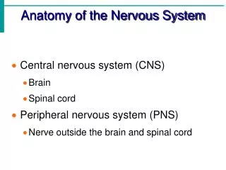
The Central Nervous System The CNS is well protected by bone, CT, and fluid Meninges– Connective tissues that surround and protect the brain and spinal cord Three layers: Dura mater, Arachnoid, Pia mater
Dura Mater – tough, fibrous outer layer; • 2 layers thick around brain with creation of dural sinuses between layers; • 1 layer around spinal cord with epidural space external Arachnoid– “spidery” web-like middle layer
Pia Mater – delicate, thin inner layer; • filum terminale - extension of pia mater extends from tip of cord to coccyx to anchor cord in place; • denticulate ligaments anchor cord laterally
Lumbar cystern Subarachnoid space – between arachnoid & pia mater; contains cerebrospinal fluid (CSF) Lumbar cistern – area of subarachnoid space below the conus medularis; site for lumbar puncture (“spinal tap”)
The Spinal Cord • Begins at foramen magnum & ends at L2 vertebral level by forming conus medularis • Has 2 thickened areas- cervical enlargement - supplies nerves to upper extremity • lumbar enlargement - supplies nerves to lower extremity • Made up of 31 spinal cord segments
Dorsal root ganglion (DRG) Dorsal root Ventral root • Each spinal cord segment has a pair of • dorsal roots with their associated dorsal root ganglia (DRG) • ventral roots
Each dorsal root contains the axons of sensory neurons • Each dorsal root ganglion contains the cell bodies of these sensory neurons • Each ventral root contains the axons of motor neurons
The dorsal & ventral roots of each segment come together at the intervertebral foramen (IVF) to form a mixed spinal nerve
Spinal Nerves • Part of the PNS • Contain both motor & sensory fibers • 31 pair of nerves – each nerve forms from union of dorsal/ventral root of spinal cord segment & exits between vertebra at IVF • 8 pair cervical spinal nerves – 1st cervical nerve exits between occipital bone & C1, 8th cervical nerve exits the IVF between C7-T1 • 12 pair thoracic spinal nerves • 5 pair lumbar nerves • 5 pair sacral nerves • 1 pair coccygeal nerves
Below the conus medularis, spinal nerves must angle downward (in the subarachnoid space) before exiting their IVF. These spinal nerves make up the cauda equina Cauda equina
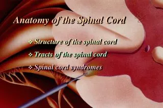
Posterior median sulcus Posterior column Posterior gray horn - sensory Central canal Lateral column Lateral gray horn (T1-L2, S2-S4) - autonomic Anterior gray horn - motor Anterior column Anterior median fissure Sectional Anatomy of the Spinal cord Gray commissure
The spinal cord has a narrow central canal surrounded by “horns” of gray matter connected by a commissure. Gray matter horns contain sensory & motor nuclei (groups of cell bodies). Gray matter is surrounded by white matter “columns” which are made up of groups of myelinated axons creating organized ascending & descending tracts.
Tracts (Motor & Sensory Pathways)(Chap. 16, p. 432-439) • Groups of axons found in the white matter columns of the spinal cord that carry specific information • Ascending tracts - carry sensory information up the spinal cord to areas of the brain (eventually terminating in cerebrum or cerebellum) • Descending tracts – carry motor information from the brain down to specific levels of the spinal cord
Ascending Tracts (Pathways) • Three major groups of pathways transmit somatic sensory information originating from receptors, up the spinal cord to the brain – • Spinothalamic tracts • Posterior column pathways • Spinocerebellar tracts
Spinothalamic tracts Anterior spinothalamic tract (ASTT) – crude touch & pressure Lateral spinothalamic tract (LSTT) – pain & temperature THALAMUS
Posterior Column Pathways • Fasciculus cuneatus & fasciculus gracilis – • conscious proprioception (joint position) • discriminitive (fine) touch (2-point discrimination, stereognosis, graphism) • vibration • pressure
Spinocerebellar Tracts Anterior spinocerebellar tract (ASCT) & Posterior spinocerebellar tract (PSCT) – • unconscious proprioception (from golgi tendon organs, muscle spindles & joint capsules) • muscle tone • balance
Descending Pathways • Carry motor signals from conscious & unconscious areas of the brain, down the spinal cord to control contraction of skeletal muscles • Corticospinal pathway (includes anterior & lateral corticospinal tracts, and corticobulbar tracts) • Medial pathways • Lateral pathways
Corticospinal (Pyramidal) Pathway • Corticobulbar tracts – voluntary control of skeletal muscles of head & neck through cranial nerves • Lateral corticospinal tracts (LCST) – voluntary control of skeletal muscles in neck & body; fibers cross in pyramidal decussation of M.O. • Anterior corticospinal tracts (ACST) - voluntary control of skeletal muscles in neck & body; fibers cross at spinal cord level in anterior commissure
Medial & Lateral Pathways Integrated with corticospinal pathways to allow for coordination of motor activity, maintenance of posture and muscle tone Medial pathways – unconscious control over neck, trunk & proximal limb muscles for gross muscle movements Lateral pathways – unconscious control over distal limb muscles for precise muscle movements Tracts include: vestibulospinal, tectospinal, reticulospinal & rubrospinal
In order for sensory information to enter the spinal cord and ascend in a sensory tract, and for motor information to get from a descending tract to reach a skeletal muscle, impulses must travel through peripheral nerves (spinal nerves & cranial nerves)
Spinal Nerves • 31 pair • Part of PNS • Formed by union of ventral (motor) root and dorsal (sensory) root
Once formed, spinal nerves will branch into Rami • Dorsal ramus – transmits sensations from skin of back & neck; provides motor control of deep muscles of back; found at all spinal nerves
Ventral ramus – provides motor control to muscles of extremities, anterior & lateral trunk; transmits sensations from all but skin of back; found at all spinal nerves
Rami communicantes(white ramus & gray ramus) – carry autonomic motor fibers (ANS) to smooth muscles & glands in ventral body cavity; transmit visceral sensations; only found at T1-L2 spinal nerves
Nerve Plexuses Adjacent ventral rami will form complex interwoven networks of nerve fibers (axons) known as a nerve plexus Four plexuses – cervical, brachial, lumbar, & sacral Emerging from each plexus will be specifically named peripheral nerves, which will contain fibers from multiple spinal cord levels
Cervical plexus (C1-C5) • Phrenic nerve (C3-C5)
Brachial plexus (C5-T1) • Axillary nerve (C5-C6) • Musculocutaneous nerve (C5-7) • Radial nerve (C5-T1) • Median nerve (C6-T1) • Ulnar nerve (C8-T1)
Lumbar plexus (T12-L4) • Femoral nerve (L2-L4) • Iliohypogastric nerve (T12-L1) • Obturator nerve (L2-L4)
Sacral plexus (L4-S4) • Sciatic nerve (L4-S3) • Tibial nerve • Common peroneal (fibular) nerve
Ventral rami from T2-T11 do not participate in a plexus. Instead they form individual intercostal nerves