Download
1 / 30
340 likes | 808 Views
Molecular Basis of Genetics and Biotechnology.

E N D
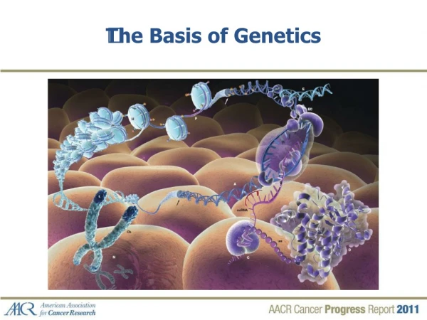
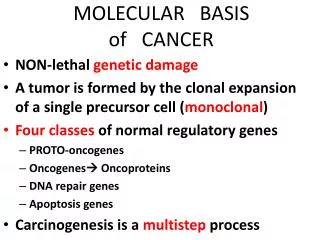
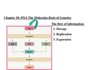
Molecular Basis of Genetics and Biotechnology The central dogma of molecular biology describes the flow of genetic information from DNARNAprotein. This flow happens through precise mechanisms, although mistake can happen during the process. Many technologies take advantage of the properties of DNA to generate novel products and tools.
Each group will be given a different experiment or insight that led to the structure and function of DNA. • Griffith experiment (2 groups) (190-191) • Avery experiment (191, additional handout) • Hershey-Chase experiment (2 groups) (192-193) • Chargaff’s observation, structure of the 4 bases (195, 196) • Wilkins and Franklin’s X-ray diffraction (196) • Watson and Crick’s DNA model and pairing between bases (196-197)
Avery experiment • Repeated Griffith experiment to see which molecule actually transformed into the harmless strain • Treated extract of heat killed smooth colonies with enzymes that broke down everything except for DNA. What happened? • Did same with enzyme that broke down DNA. What happened?
Watson and Crick Model • What did they know: DNA was a molecule involved in genetic information • What did they not know: The structure of the molecule • What evidence did they use? • X-Ray diffraction: Scattered pattern of DNA X-rays on film produced by Rosalind Franklin (showed coiled strand and angle of stands) • Chargaff’s rule
What is the basic unit (monomer) of a nucleic acid? What are the 3 subunits of a nucleotide. Draw them. Which of these subunits are the same in all nucleic acids? What are the 4 bases of DNA? What is the function of DNA, can it leave the nucleus? Reading quiz – Read through WB 34. I will stamp SG 1 for full credit today only. I will also stamp SG 3 on Thursday for full credit (if not done)
Analysis question 2 • Draw a labeled diagram of your DNA molecule. Keep the illustration in a straight “ladder” form as seen in figure 5. Do not attempt to draw the helical shape. Identify each nitrogen base. • Do this on the back of WB 34
Structure of DNA • Double stranded helix • Deoxyribose sugar • Phosphate group • 4 nitrogen bases • Guanine : Cytosine • Adenine : Thymine • Hydrogen bonds connect bases
DNA replication • Each strand is complementary • Replication steps • DNA Helicase (enzyme) “unzips” the DNA strand, breaking the hydrogen bonds • DNA polymerase adds complementary bases to each strand (5’ to 3’) • Sugar phosphate links extend the chain and DNA polymerase “proof reads” new strand • DNA ligase seals fragements together
Person closest to the molecular modeling kit Read through WB 35 Rebuild your double helix exactly as it was on Monday/Tuesday. (You can use your instructions). Make sure you have the correct base sequence and base pairing. THIS IS YOUR READING QUIZ! Everyone else at the table Read through WB 35 and define the following terms. 1. Anticodon 2. Codon 3. Nucleotide 4. Ribosome 5. Transcription Reading quiz
DNA model Check with model up front 1. Anticodon: 3 nucleotides on a tRNA complementary to the codon 2. Codon: 3 nucleotides of an mRNA that codes for an amino acid 3. Nucleotide: Monomer of a nucleic acid consisting of a sugar, base, and phosphate 4. Ribosome: Organelle which is the site of protein synthesis 5. Transcription: Process of producing an mRNA molecule from a DNA strand Reading quiz answers
Complete letters C-F using the DNA molecule just made • Answer these questions on the back of WB 35 (yep, we’re conserving paper) • 3. A partial DNA stand has the following sequence: CACTTGCAC. What would be the complementary mRNA sequence • 4. How can protein be synthesized in the cytoplasm of a cell when DNA is contained in the nucleus?
Structure of RNA • Can leave the nucleus • Single stranded • Ribose sugar • Phosphate group • 4 nitrogen bases • Guanine : Cytosine • Adenine : Uracil
Transcription • The process of copying part of a DNA strand into a complementary RNA strand, so the information can be taken outside of the nucleus without affecting the DNA molecule
Reading quiz. Use study guide and WB 34-35. Staple exam to back of test corrections and turn in. • Complete the following chart
Complete letter G • Answer the following questions on the back of WB 36 • 5. You have the following mRNA sequence GUGAACGUG. What would be the anticodon base sequence on the corresponding tRNAs? Circle each codon and anticodon. • 6. Using a venn diagram, compare and contrast codons to anticodons (structure? Function? Location?)
English code Genetic code • Has a four letter alphabet • There are 20 different amino acids (protein building blocks) • How many letters codes for each amino acid? • Codon: 3 letter combination of mRNA strand
tRNA • Anticodon (complementary to mRNA) • Attached amino acid • Codon: AAA, then anticodon is ____
Left table of your group. Put together the mRNA molecule produced in letter F (hint, look at the diagram on WB 40 and think about which sugar to use) Right table of your group. Put together the tRNA molecules produced in letter G. Add appropriate amino acid (black) to each tRNA Reading quiz
Translation • Goal of genetic code is to produce a protein. How does this happen? • 1) Transcription produces mRNA strand • 2) mRNA binds with ribosome • 3) tRNA anticodon binds with mRNA • 4) Amino acids attached to tRNA bond together
Hookin’ up amino acids • mRNA and first tRNA bind to ribosome (but not each other yet). This is always the anti codon UAC (amino acid – methionine) • Ribosome scans mRNA and keeps first codon sequence (AUG) at the P site. Anticodon hydrogen bonds with 1st codon • Anticodon complementary to the 2nd codon hydrogen bonds to it at the A site. • Amino acid of 1st tRNA detaches and forms peptide bonds with amino acid of 2nd tRNA • mRNA and 2nd tRNA move to the left as the 1st tRNA leaves ribosome • When a “stop codon” is reached polypeptide chain leaves, other parts dissemble • Polypeptide may be modified (where?) before it is a functional protein
Write response in notebook • Use the genetic code (on your desk) to find the amino acid sequence coded by the mRNA sequence AUGAAGUUU • Use the genetic code (on your desk) to find the amino acid sequence coded by the DNA sequence TATCATGCC
Mutations • Point • Frameshift • THE CAT ATE THE RAT • THC ATA TET HER AT • Mutations constantly happen (about 1 in every 1000 bases) and are important for variation Severity depends on location and number of mutations
Gene regulation • All cells in an organism have the same genes • Do all cells look the same and do the same job? Why? • How is this possible?