Download
1 / 44
540 likes | 1.16k Views
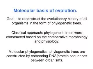
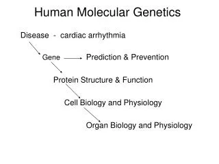
Molecular Basis of Genetic Diseases and Tools of Human Molecular Genetics. Ömer Faruk Bayrak. Genetic Diseases in Humans. Role of Genes in Human Disease. Struck by lightning. 100% Environmental. Most diseases -> phenotypes result from the interaction between genes and the environment

E N D
Molecular Basis of Genetic DiseasesandTools of Human Molecular Genetics Ömer Faruk Bayrak
Genetic Diseases in Humans Role of Genes in Human Disease Struck by lightning 100% Environmental • Most diseases -> phenotypes result from the interaction between genes and the environment • Some phenotypes are primarily genetically determined • Achondroplasia (-> dwarfism) • Other phenotypes require genetic and environmental factors • Mental retardation in persons with PKU (polyketonuria) • Some phenotypes result primarily from the environment or chance • Lead poisoning Infection Weight Cancer Diabetes Height 100% Genetic Down syndrome, achondroplasia
Principles of moleculardisease Molecularreason of a geneticdisease is a mutation. Thismutationeitherinheritedoracquired. Thebiochemicalgenetic is study of phenotype at thelevel of proteins, biochemistryandmetabolism.
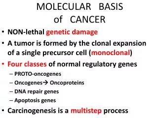
Principles of moleculardisease Effects of Mutations, • Lossof function (αthalasemia) • Gaina function (Achondroplasia) • Acquisionof a novelpropertybymutant protein. (SickleCell Anemia) • Expressionof a gene at thewrong time orwrongplace. (most of cancers)
Genetic Diseases in Humans Types of Genetic Disorders: -> Chromosomes and chromosome abnormalities (Down Syndrome) -> Single gene disorders (Haemophilia, sickle cell anaemia) -> Polygenic Disorders (Cancer)
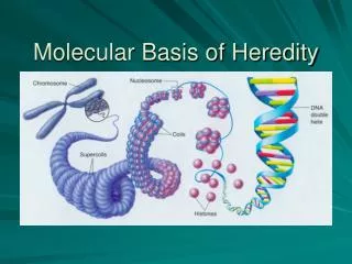
Genetic Diseases in Humans Chromosomal disorders • Addition or deletion of entire chromosomes or parts of chromosomes • Typically more than 1 gene involved • 1% of paediatric admissions and 2.5% of childhood deaths • Classic example is trisomy 21 - Down syndrome KARYOTYPE
Genetic Diseases in Humans Single gene disorders • Single mutant gene has a large effect on the patient • Transmitted in a Mendelian fashion • Autosomal dominant, autosomal recessive, X-linked, Y-linked • Osteogenesis imperfecta - autosomal dominant • Sickle cell anaemia - autosomal recessive • Haemophilia - X-linked
Genetic Diseases in Humans Single gene disorders Neonatal fractures typical of osteogenesis imperfecta, an autosomal dominant disease caused by rare mutations in the type I collagen genes COL1A1 and COL1A2 A famous carrier of haemophilia A, an X-linked disease caused by mutation in the factor VIII gene Sickle cell anaemia, an autosomal recessive disease caused by mutation in the β-globin gene
Genetic Diseases in Humans Polygenic disorders • The most common yet still the least understood of human genetic diseases • Result from an interaction of multiple genes, each with a minor effect • The susceptibility alleles are common • Type I and type II diabetes, autism, osteoarthritis, cancer
Monogenic Disorders • Involve single mutant genes • Classification: (1) autosomal dominant - clinically evident if one chromosome affected (heterozygote) • e.g., Familial hypercholesterolemia (2) autosomal recessive - both chromosomes must be affected (homozygous) • e.g., Sickle cell anemia (3) X-linked - mutation present on X chromosome • females may be either heterozygous or homozygous for affected gene • males affected if they inherit mutant gene • e.g., Duchenne muscular dystrophy
Multifactorial Disorders • Interplay of number of genes and environmental factors • pattern of inheritance does not conform to classic Mendelian genetic principles • due to complex genetics, harder to identify affected genes; thus, less is known about this category of disease • e.g., Essential hypertension
The Various Types of Heterogeneity Associated with Genetic Disease
Some examples of Classes of Proteins Associated with Monogenic Diseases • 1-Transport andstorage • İnterorgan Hemoglobin (thalassemias)(AR) • İntracellular transport (copper transport prot. menkessyndr. (AR) • Epitelmembr. Cysticfibrosis (CFRT AR) 2-Enzyme defects • Amino acids - PKU(phenylalaninehydroxilase AR) • Complexlipid-Tay sachs(Hexosaminidase A AR) • Purines-immundeficiency( Adenosinedeaminidase AR) • CarbohydratesGalactose 1 phosphateuridyltransferase
Some examples of Classes of Proteins Associated with Monogenic Diseases 3-Structure of cells and organs • Duschene muscular distrophy(dystrophin XR) 4-Control of growth anddifferantiation • Tumorsuppressors, • RB gene products (AR), • oncogenes(AD). 5-Intracellular metabolism and comunication • Growth gormon(dwarfizm,AR),insulin(AD) • Familialhypercholesterolemia(LDL receptor).
Tools of Human Molecular Genetics PCR • PCR was first conceived in 1983 by Kary Mullis, a molecular biologist who received a Nobel Prize for the discovery 10 years later • A PCR (Polymerase Chain Reaction) is performed in order to make a large number of copies of a gene. Otherwise, the quantity of DNA is insufficient and cannot be used for other methods such as sequencing. • A PCR is performed on an automated cycler, which heats and cools the tubes with the reaction mixture in a very short time. • Performed for 30-40 cycles, in three major steps: 1)denaturation, 2)annealing, and 3)extension.
1) Denaturationat 94°C : • During the denaturation, the double strand melts open to single stranded DNA, all enzymatic reactions halt. 2) Annealingat 50-60°C : • The primers are freely moving due to Brownian motion. Ionic bonds are constantly formed and broken between the single stranded primer and the single stranded template. • Primers that fit exactly will have stable bonds that last longer. The polymerase attaches onto a piece of double stranded DNA (which is template and primer), and starts copying the template. Once there are a few bases built in, the ionic bond is so strong between the template and the primer, that it does not breakanymore. 3) Extensionat 72°C : • This temperature is ideal for the polymerase. The primers, which have a few bases built in, already have a stronger ionic attraction to the template than the forces breaking these attractions. • Primers that are on positions with no exact match, loosen their bonds again (because of the higher temperature) and do not extend the fragment. The bases (complementary to the template) are coupled to the primer on the 3' side (the polymerase adds dNTP's from 5' to 3', reading the template from 3' to 5' side, bases are added complementary to the template)
The ladder is a mixture of fragments with known size to compare with the PCR fragments. Notice that the distance between the different fragments of the ladder is logarithmic. Lane 1 : PCR fragment is approximately 1850 bases long. Lane 2 and 4 : the fragments are approximately 800 bases long. Lane 3 : no product is formed, so the PCR failed. Lane 5 : multiple bands are formed because one of the primers fits on different places.
Applications of PCR: • 1) Diagnosis of Disease: Linkage analysis, detection of mutant alleles, diagnosing infectious agents, epidemiological studies • 2) Forensics: paternity testing, DNA typing for identification, criminal investigations. • 3)Recombinat DNA engineering • 4) DNA sequence determination • 5) new gene isolation • 6) Anthropological studies: population genetics, migration studies. • 7) Evolution studies • If you need to look at 100 genes is PCR a good approach?
RT-PCR • An RT-PCR (Reverse transcriptase-polymerase chain reaction) is a highly sensitive technique for the detection and quantitation of mRNA (messenger RNA). • The technique consists of two parts: 1) The synthesis of cDNA (complementary DNA) from RNA by reverse transcription (RT) 2) The amplification of a specific cDNA by PCR. Compared to Northern blot analysis and RNase protection assay used to quantify mRNA, RT-PCR can be used from much smaller samples. It is sensitive enough to enable quantitation of RNA from a single cell. • Real-time RT-PCR is the method of choice for quantitating changes in gene expression. Furthermore, real-time RT-PCR is the preferred method for validating results obtained from array analyses and other techniques that evaluate gene expression changes.
Real-time PCR advantages * not influenced by non-specific amplification * amplification can be monitored real-time * no post-PCR processing of products (high throughput, low contamination risk) * ultra-rapid cycling (30 minutes to 2 hours) * wider dynamic range of up to 1010-fold * requirement of 1000-fold less RNA than conventional assays (3 picogram = one genome equivalent) * detection is capable down to a 2-fold change * confirmation of specific amplification by melting curve analysis * most specific, sensitive and reproducible * not much more expensive than conventional PCR (except equipment cost)
Microarray • DNA microarrays allow researchers to analyze the expression of thousands of genes simultaneously. • DNA microarrays contain thousands of individual gene sequences in microscopic spots of ≈1-kb DNA sequences representing thousands of genes bound to the surface of glass microscope slides. • Provide a means for analyzing gene expression patterns on a genomic scale. • Provides a medium for matching known and unknown DNA samples based on base-pairing rules and automating the process of identification.
Microarray • Applications • Gene discovery • Disease diagnosis • Drugs and toxicological research: The goal of pharmacogenomics is to find correlations between therapeutic responses to drugs and the genetic profiles of patients. • Expression screening. The focus of most current microarray-based studies is the monitoring of RNA expression levels which can be done by using either cDNA clone microarrays or gene-specific oligonucleotide microarray • Screening of DNA variation. There is also huge potential for assaying in drug development and patient susceptibility, as well as for mutations in known disease genes such as cardiovascular disease and cancer as seen in the case of the breast cancer susceptibility gene, BRCA1. • In addition, there have been vigorous efforts to identify and catalog • human single nucleotide polymorphism (SNP) markers.
Gene Expression Analysis Technologies >10,000 Real-time PCR 100 Low-density Arrays Number of samples 10 High-density Arrays RPA Northern SAGE 1 2-10 100-1000 >10,000 Number of genes To consider: Ratio samples / genes, needs for accuracy
Comparison of Quantitative Assays (RNA/DNA): Sensitivity Dynamic Range Real-Time PCR Amplicor/TMA NASBA bDNA XPLORE Microarrays RPA Northern 100 101 102 103 104 105 106 107 108 108 107 106 105 104 103 102 101 100 NASBA: nucleic acid seq based amplification bDNA: branched DNA assay Xplore: based on Invader technology TMA: transcription mediated amplification RPA: RNAse protection assay Advantage: Real-Time PCR
Southern Blot • Southern Blotting (named after Ed Southern, the inventor) is the detection of specific sequences of DNA on a gel by hybridisation with a labelled DNA probe. • DNA is first transferred out of a gel by capilliarity (the "blot") to a thin membrane which can be incubated with a probe and washed. • By hybridising at different temperatures, and washing to different ionic strengths ("stringencies") it is possible to tune the process to pick up sequences that are either similar, or exactly identical, to the probe.
Southern Blot • Applications: • 1)To confirm the presence of a gene, often in conjunction with PCR. • 2)To test for the presence of a specific allele of a gene (i.e. human disease genetics). • 3)To estimate gene complexity, before you have the gene sequence. • 4) To detect Restriction Fragment Length Polymorphism (RFLP) and Variable Number of Tandem Repeat Polymorphism (VNTR). The latter is the basis of DNA fingerprinting.
Northern Blots • Northern blots are similar to Southern, except that RNA from different tissues is run out on a gel, and probed with a DNA or RNA probe corresponding to a particular gene. • Northern blotting is used for detecting and quantitation of RNA fragments, instead of DNA fragments. The technique is exactly like Southern Blotting. It is called "Northern" simply because it is similar to "Southern", not because it was invented by a person named "Northern". • RNA samples are first separated by size via electrophoresis in an agarose gel under denaturing conditions. The RNA is then transferred to a membrane, crosslinked and hybridized with a labeled probe.
Western Blot • Western blot analysis can detect oneprotein in a mixture of any number of proteins while giving you information about the size of the protein. • Allows investigators to determine with a specific primary antibody, the relative amounts of the protein present in different samples. • Western blots are analogous to Northern and Southern, except that proteins are run out in an SDS polyacrylamide gel, and are detected with specific antibodies. • In clinical settings, Western Blotting is routinely used to confirm serious diagnosis suggested by ELISA such as HIV seroconversion
Nucleic Acid Hybridization • The Basic Process of Binding a Single Strand of Nucleic Acid (DNA or RNA) to Its Complementary Strand Is Called Nucleic Acid Hybridization. • Double-stranded DNA Can Be Denatured by Agents Such As Heat or High PH. When Denatured, the Two Strands Separate Into Single Strands and Diffuse Away From Each Other. If Conditions Are Then (Slowly) Reversed (Lower the Temperature or Return the PH to Neutrality) Then the DNA Will renature. • If the Temperature Is Slowly Decreased, Then Each Strand of DNA Will Find Its Corresponding Mate: the Complementary Strands of the DNA Will Anneal and Re-form the Double Strand With Correct Watson-crick If a Radioactively Labeled Probe Corresponding to a Part of the Sequence of One of the Fragment Is Included in the renaturation Mixture, It Will Participate in the renaturation, Finding and Annealing to Its Complementary Partner
Nucleic Acid Hybridization Probe present No probe
A C C C T G C G
FISH • Fluorescence In-Situ Hybridization is a method used to identify specific parts of a chromosome. For example, if you know the sequence of a certain gene, but you don't know on which chromosome the gene is located, you can use FISH to identify the chromosome in question and the exact location of the gene. • If you suspect that there has been a translocation in a chromosome, you can use a probe that spans the site of breakage/translocation. If there has been no translocation at that point, you will see one signal, since the probe hybridizes to one place on the chromosome. If, however, there has been a translocation, you will see two signals, since the probe can hybridize to both ends of the translocation point. • To use FISH efficiently, you have to know what you're looking for, i.e. you usually suspect a particular defect, based on the appearance of certain chromosomes, etc.
FISH Method: • Make a probe complementary to the known sequence. When making the probe, label it with a fluorescent marker, e.g. digoxigenin, by incorporating nucleotides that have the marker attached to them. • Put the chromosomes on a microscope slide and denature them. • Denature the probe and add it to the microscope slide, letting the probe hybridize to its complementary site. • Wash off the excess probe and look at the chromosomes in a fluorescence microscope. The probe will show as one or more fluorescent signals in the microscope, depending on how many sites it can hybridize to.
FISH Applications • Diagnosis in clinical and cancer cytogenetics. • Interspecies studies of evolutionary divergence. • Analysis of aberrations in animal models of human diseases.