Download
1 / 38
380 likes | 574 Views
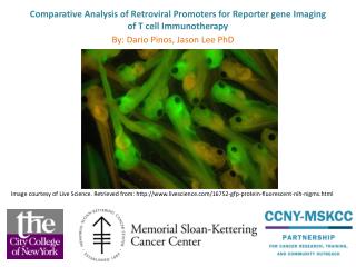
HSV1 Thymidine Kinase Reporter Gene Imaging. 싸이클로트론 응용연구실 원자력병원. Methods for the measurement of gene expression. Conventional measurement of Gene expression. Southern, Northern , and Western blot and in situ hybridization. Reporter gene

E N D
HSV1 Thymidine Kinase Reporter Gene Imaging 싸이클로트론 응용연구실 원자력병원
Methods for the measurement of gene expression Conventional measurement of Gene expression Southern, Northern , and Western blot and in situ hybridization Reporter gene easier, less expensive, more accurate & quantitative than standard hybridization technique Reporter gene and In Vivo imaging of gene Imagene Jean-Luc C. Urbain ; JNM Vol.42, No.1, Jan , p106-109
Widely Used Reporter Gene Systems • b-galactosidase system b-galactosidase • b-glucuronidase system b-glucuronidase • CAT (CM acetyl transferase) system CM acetyl transferase • SEAP (secreted alk-phosphatase) system alkaline phosphatase • Firefly luciferase system luciferase • GFP (green fluorescent protein) system green fluorescent protein • HSV1-TK system
Principle of Conventional Reporter Assays Reporter gene Reporter Proteins Histological staining mRNA Transfection DNA Biochemical assay Photon detection
reporter gene system for in vivo imaging of transgene • HSV1-tk : various purine & pyrimidine substrates : development of radiolabeled analogs Nuclear Imaging Reporter gene Radiolabeled substrate trapped Reporter Enzymes Transfection Reporter Receptors Radiolabeled ligand • transmembrane receptor - receptor ligand ex) Dopaminergic type2 receptor - 18F-fluoroethylspiperone
HSV1-TK System HSV1-tk (Herpes Simplex Virus type 1 – thymidine kinase) relaxed substrates specificity substrates : thymidine, purine pentoside, wide diversity of nucleoside analogues Synthetic purine / pyrimidine nucleoside derivatives Monophosphate form Diphosphate form Triphosphate form ( acyclovir, ganciclovir ) Other cellular kinase HSV1-tk Guanylate kinase
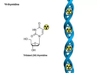
Radiolabeled susbtrates for HSV1-tk reporter system Radiolabel • H-3, C-14 : Autoradiography • I-131, I-123 : SPECT • I-124, F-18 : PET Uracil nucleoside derivatives (higher enzyme affinity) Acycloguanosine derivatives (F-18 labeling possible) • FIAU • IVDU • IVFRU • IVFAU • IVAU • FACV • FGCV • FPCV
Uracil nucleoside 계 Acycloguanosine 계
Receptor-Ligand Reporter System Dopamine 2 Receptor (D2R) : 3-(2’-fiuoroethyl) spiperone (FESP) reporter system Membranous receptor - labeled cognate receptor ligand Bicistronic adenoviral vector carrying human D2R and somatostatin type 2 receptor(hSSTr2) Na+ /I symporter (NIS) gene reporting & therapeutic system Not only radioiodine therapy but also transfected tumor localization and gene therapy assays
Prerequisites for using a reporter gene imaging Reporter Gene • Does not exceed size constraints of the vector • Expression of reporter gene is stable and reflect expression of targeted gene • The endogeneous gene is expressed only in low levels • The vector does not induce significant host immune response • Can be dual inserted to reflect expression of gene of interest Reporter Protein • Does not induce a significant immune response ( not foreign ) • Is retained in the cells that they have been expressed • Is stable and do not produce significant confounding metabolites • Does not cause significant biological effects • Endogeneous activity and Concentration is low & absent from targeted cells
Reporter Probe • Non-immunogenic & no or minimal side effects • Labeling method : single photon or positron suitable for SPECT and PET imaging • Can be radio-labeled with high specific activity • Radio-labeled reporter probe retains chemical activity • Stable in the blood & cleared rapidly from blood stream • Rapidly metabolized by reporter gene product & effectively trapped in transduced cell • Specifically retained by transfected cell & reflect activity of transgene expression • Reporter substrate readily bind to gene product as receptor - ligand • / must be able to cross cell membrane as HSV1-tk system • Radio-signal correlates with transgene expression level
암 유전자 치료 1. 암세포 : 유전적 변이에 기인 - 암유전자 (oncogene), 종양억제유전자 (tumor suppressor gene) 2. 암세포에서 발견되는 유전자이상을 교정, 조절하는 치료법 - 유전자 표지(genetic marking) - 암 백신(cancer vaccination) - 암 유전자 발현 억제 - 종양억제유전자 회복 - 자살유전자 (suicide gene)
HSV-1tk 유전자 영상 원리 HSV-1tk 유전자가 유입(transfection)된 세포는 thymidine kinase효소를 생산하므로 이 효소의 기질 (substrate, 치료는 ganciclovior)에 감마카메라로 영상을 얻을 수 있는 방사성동위원소를 표지하여 유전자 영상을 얻고, 유전자 치료의 성공여부를 조기판정 본 연구는 HSV-1tk 유전자 영상을 얻기 위하여 두 가지 약제(IVDU, IVFRU)를 합성하고, 방사성동위원소 표지하여 유전자 주입 동물모델에서 성공적인 유전자 영상을 얻었다.
Accumulation & Cytotoxicity HSV-tk Death mRNA HSV-tk gene Ganciclovir (GCV) Live HSV-1tk Gene Therapy system
Accumulation & Cytotoxicity HSV-tk Death mRNA HSV-tk gene Ganciclovir (GCV) Live HSV-1tk Gene Therapy system
Accumulation HSV-tk Image mRNA Radiolabeled HSV-tk substrate HSV-tk gene HSV-1tk Gene Imaging system
O I HN O N O OH OH R HSV-1tk 유전자 영상 제재 Nucleoside derivatives . Antiviral agent . Pyrimidine Nucleoside Analogs . Herpes Viral Infections . Imaging Reporter Gene Expression of Viral thymidine kinase R = H, (E)-5-(2-Iodovinyl)-2’- deoxyuridine (IVDU)R = F, (E)-5-(2-Iodovinyl)-2’- fluoro-2’-deoxyuridine (IVFRU)
O O * I S i M e 3 H N H N H O N 1 2 5 1 3 1 O N a I o r N a I H O N O O O I C l , 2 0 % A c O H / C H C N X = H I V D U 3 r . t , 2 0 m i n X = F I V F R U O H X O H X 동위원소 표지 순수분리 HPLC column : mBondapak - C18 Solvent : CH3CN and D.W. , Gradient Flow Rate : 1ml/min Radiochemical yield : >98%
O * I H N H O N O O O H X 연구방법 Cell line : MCA( rat Morris hepatoma Cell Line) and MCA-tk(HSV-1tk gene transfected (MCA Cell Line) HSV-1tk substrate : [125I],[131I] IVDU and IVFRU *I = 125I or 131I X = H IVDU X = F IVFRU
방사성 요오드 표지법 : Radioiodination of Substrates Trimethyl-Silyl-Vinyl-2’-fluoro-2’-deoxyuridine(SiMe3-VFRU) 50 ml(5 mg/ml) & Trimethyl-Silyl-Vinyl-2’-deoxyuridine(SiMe3-VDU) 1N HCI 100 ml 30% H2O2 50 ml Na125I 18.5MBq 10minutes reaction in Room Temp. Saturated NaSO3 100 ml Saturated NaHCO3 1ml
정제 및 분리 : HPLC analysis Column ( mBondapak C18, 3.9 x 300mm Waters ) Pump control : Gradient Flow rate : 1 ml/min Solvent : A = CH3CN B = D.W. 1-5 min 10% A : 90% B 5-10 min 40% A : 60% B 10-25 min 40% A : 60% B 25-30 min 10% A : 90% B Run time : 30 minutes Detector : UV (Waters 486) = 254nm , RI(Raytest)
800 160 IVFRU IVDU 700 140 120 600 100 500 CPM (x1,000) CPM (x1,000) 400 80 300 60 40 200 20 100 0 0 5 10 15 20 25 30 Time Radiochromatograms of IVDU and IVFRU
세포섭취율 : In Vitro Uptake in MCA & MCA-tk Cell Lines Cell Lines (1 x 10⁶ / flask) : MCA ( control ) MCA-tk ( HSV-tk gene tranduced ) Culture Conditions : 37oC , CO2 Incubator Substrates : 125IVDU, 125IVFRU (185KBq) Incubation Period : 15, 30, 60, 120, 240, 480 minutes Counting : Gamma Counter (WALLAC wizard)
125IVDU 125IVFRU 50 12 10 40 8 30 MCA MCA % injected dose MCA-tk 6 MCA-tk 20 4 10 2 0 0 0 100 200 300 400 500 600 0 100 200 300 400 500 600 Incubation time(min) Incubation time(min) 세포섭취율 : In Vitro Uptake in MCA & MCA-tk Cell Lines
유전자 주입 종양 이식 백서에서 체내 분포 : Biodistribution of 131IVDU and 125IVFRU in Tumor bearing Rats Tumor cell (1 x 10⁷ /100ml ) injected into both thigh MCA : Right thigh I.M. MCA-tk : Left thigh I.M. Two weeks later 131IVDU( 370KBq/ 100ml ) , 125IVFRU ( 370KBq/ 100ml ) Simultaneously injected I.V. into tail vein Time : After injection, 1hr / 4hr / 24hr Counting : Gamma Counter in each energy windows
혈중제거율 : Blood Clearance in Rats 4 IVFRU 125 3 IVDU 125 ID%/gm 2 1 0 0 20 40 60 80 100 120 Minutes
5 1hr 4hr 4 24hr 3 % Injected Dose 2 1 0 Blood Lung Femur Liver Kidney Tu(TK) Stomach Spleen Thyroid Muscle Intestine Tu(MCA) 체내분포 및 동태 : Biodistribution of 131IVDU
HPLC analysis of 2 hrs Urine 70 78.8% IVFRU 60 IVDU 50 Tot % 40 76.1% 30 20 18.1% 19.8 % 10 0 0 5 10 15 Fraction No.
1 hour 3 4 hour 2.5 24 hour 2 % ID/g 1.5 1 0.5 0 MCA Liver Lung Blood Femur Spleen Kidney MCA-tk Muscle Thyroid Stomach Intestine 체내분포 및 동태 : Biodistribution of 131IVDU
1 hour 2 4 hour 24 hour 1.5 %ID/g 1 0.5 0 MCA Liver Lung Blood Femur Spleen Kidney MCA-tk Muscle Thyroid Stomach Intestine 체내분포 및 동태 : Biodistribution of 125IVFRU
II-5. In Vivo Image of 123IVDU and 123IVFRU In tumor bearing Rats Model 1 : MCA-tk(HSV-tk gene transduced) cells (1 x 10⁷ /100 ml ) injected S.C. into Right thigh Model 2 : Tumor cells (1 x 10⁷ /100 ml ) injected S.C. into both thigh MCA : Right MCA- tk : Left Substrates : 123IVDU, 123IVFRU( 74MBq / 200 ml ) injected I.V into tail vein In Hepatocellular Carcinoma bearing Rats Model : MCA- tk cells (1 x 10⁷ /100 ml ) injected S.C. into Shoulder of Rat Establishd tumor mass implanted into liver using intra-hepatic tumor injection Substrates : 123IVDU( 74MBq / 200 ml ), 123IVFRU( 74MBq / 200 ml ) Gamma camera image : After injection, 2hr / 24hr Sophy Camera DSX rectangular - 300,000 counts collect
Rat Orthotopic Hepatoma Model of MCA-RH7777 Intrahepatic Tumor Implantation (IHTI) Subcutaneous Implantation Transabdominal Ultrasonogram
TK TK Thyroid MCA TK MCA Monitoring of Gene Therapy : 131IVDU
MCA-tk MCA-tk 유전자 영상 (피하종양이식모델) 123IVDU 123IVFRU 2hr 24hr 2hr 24hr
HCC HCC HCC HCC 유전자 영상 (간종양이식모델) 123IVDU 123IVFRU 2hr 2hr section section 24hr 24hr
MCA MCA MCA-tk MCA-tk 125IVDU 125IVDU 125IVDU 125IVDU (1hr) (4hr) (1hr) (4hr) Cell/Media 0.5 0.5 17.2 44.8 Nuclei 16.1 14.7 27.8 55.2 Mitochondria 9.1 8.6 47.7 29.8 Microsome 4.2 4.8 7.5 6.8 Cytosol 70.5 72.0 16.9 8.2 Table 1. Intracellular localization of 125IVDU at 1hr and 4hr in MCA and MCA-tk cells
MCA MCA-tk MCA-tk 125IVDU 125IVDU 125IUdR Cell/Media 0.5 44.8 1.8 Nuclei 14.7 55.2 54.3 Mitochondria 8.6 29.8 8.9 Microsome 4.8 8.2 3.5 Cytosol 71.9 6.8 33.3 Table 2. Intracellular localization of 125IUdR and 125IVDU at 4 hr in MCA and MCA-tk cells
결 론 세포섭취율 (In Vitro Uptake) : IVFRU 116 - fold, IVDU 37 - fold in 480 min IVFRU > IVDU 체내분포 (In Biodistribution): Both IVDU and IVFRU present similar pattern 유전자영상 (In Vivo Image) Model 1 : Both IVDU and IVFRU showed defined image HCC Model : IVFRU is selectively localized in HCC 결론: IVDU and IVFRU could be useful for gene monitoring agents