Patient Monitors
520 likes | 1.02k Views
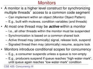
Patient Monitors. R1 김영석. “vigilance”. The motto of the American Society of Anesthesiologists. CARDIAC MONITORS Arterial Blood Pressure Electrocardiography Central Venous Catheterization Pulmonary Artery Catheterization Cardiac Output

Patient Monitors
E N D
Presentation Transcript
Patient Monitors R1 김영석
“vigilance” The motto of the American Society of Anesthesiologists
CARDIAC MONITORS Arterial Blood Pressure Electrocardiography Central Venous Catheterization Pulmonary Artery Catheterization Cardiac Output PULMONARY MONITORS Precordial & Esophageal Stethoscopes Pulse Oximetry Capnography Anesthetic Gas Analysis NEUROLOGICAL SYSTEM MONITORS Electroencephalography Evoked Potentials MISCELLANEOUS MONITORS Temperature Urinary Output Peripheral Nerve Stimulation
Arterial Blood Pressure Pulse pressure : systolic pressures 와 diastolic pressures 의차이 Mean Arterial Pressure : The time-weighted average of arterial pressures during a pulse cycle : MAP = {(SBP) + 2 (DBP)} / 3 측정 부위에 가장 큰 영향을 받음 심장의 높이와의 차이에 따른 중력의 영향 : 1cm = 0.74mm Hg
Noninvasive Arterial Blood Pressure Monitoring Indications : 모든 마취 Contraindications : in extremities with vascular abnormalities or with intravenous lines Techniques a. Palpation b. Doppler Probe c. Auscultation d. Oscillometry e. Arterial Tonometry
Noninvasive Arterial Blood Pressure Monitoring Palpation : cuff로 막혀있던 맥박이 다시 촉지될때의 cuff pressure를 측정 : 실제 systolic pressure보다 낮게 측정 되는 경향 : diastolic pressures와 MAP의 측정 불가능 Doppler Probe : 손가락 대신 doppler probe를 사용 red blood cell 의 움직임을 측정 : obese patients, pediatric patients, patients in shock에 유용 : systolic pressures 만 측정 가능 Auscultation : Kortokoff sounds를 청진 : cuff pressure를 systolic과 diastolic pressure사이에 두었을때의 turbulent flow
Noninvasive Arterial Blood Pressure Monitoring Oscillometry : arterial pulsations 은 진동을 유발 : cuff pressure가 systolic pressure 아래일때 진동은 cuff전체로 전달 : 진동은 MAP에서 가장 높음 : 부정맥이 있을경우 부정확 할 수 있음 : 가장 선호되는 noninvasive blood pressure monitors
Noninvasive Arterial Blood Pressure Monitoring Arterial Tonometry : pressure transducers를 동맥이 지나는 피부위에 위치 : invasive arterial blood pressure와 유사한 waveform 측정가능 : 지속적인 감시가 가능 : 장치의 움직임에 민감해 잦은 calibration의 필요
Noninvasive Arterial Blood Pressure Monitoring Clinical Considerations : 마취중 vital organ으로의 충분한 산소의 전달이 유지되어야 함 : arterial blood flow는 장기로의 혈류량을 반영 : but, 정확한 측정값으로 볼 수는 없음 ( ∵ Flow = Pressure / Resistance ) : 적절한 cuff size가 중요 : 자동혈압기의 너무 잦은 측정은 nerve palsy, 정맥으로 주입한 fluid의 extensive extravasation을 유발
Invasive Arterial Blood Pressure Monitoring Indications - induced hypotension - wide blood pressure swings가 예상되는 경우 - 정확한 blood pressure가 필요한 end-organ disease - 반복적인 arterial blood gas analyses가 필요한 경우 Contraindications - collateral blood flow가 없는 경우 - preexisting vascular insufficiency (eg, Raynaud’s phenomenon)
Invasive Arterial Blood Pressure Monitoring Selection of Artery for Cannulation a. Radial artery (most common) - superficial한 위치 - collateral flow의 존재 - 5%에서 불충분한 collateral blood flow - Allen’s test b. Ulnar artery - deeper and more tortuous - 잘 선택되지 않음
Invasive Arterial Blood Pressure Monitoring Selection of Artery for Cannulation c. Brachial artery - less waveform distortion - kinking의 가능성 d. Femoral artery - excellent access - infection - arterial thrombosis e. Dorsalis pedis and Posterior tibial arteries - most distorted waveforms f. axillary artery - 주변 axillary nerve plexus damage ( ∵ hematao or traumatic cannulation) - air 나 thrombi의 cerebral circulation 으로 유입 가능성
Invasive Arterial Blood Pressure Monitoring Technique of Radial artery cannulation - the pressure-tubing-transducer system과 heparinized saline 준비 - 손목을 supination and extension 시켜 고정 - radial pulse 촉진 (with 2nd and 3rd fingers) - skin preparation (with a bactericidal agent) - local anesthetic 주사(with 25G needle) - 45도 각도로 촉지부위를 향해 전진 (with 20 or 22G catheter) - Blood flashback이 확인되면 needle을 30도로 낮춘후 1-2mm 더 전진 - catheter를 vessel lumen내로 삽입 : cathter를 spinning 시키면서 삽입 - 3rd and 4th fingers로 proximal쪽에 압력을 가해 실혈을 막음 - tubing과 catheter연결 - water-proof tape등으로 고정
Invasive Arterial Blood Pressure Monitoring Complications - hematoma - bleeding - vasospasm - arterial thrombosis - embolization of air bubbles or thrombi - necrosis of skin - nerve damage - infection - loss of digits - unintentional intraarterial drug injection
Invasive Arterial Blood Pressure Monitoring Complications의 증가요인 - prolonged cannulation - hyperlipidemia - 여러 번의 삽입 시도 - female - extracorporeal circulation - vasopressors 의 사용
Invasive Arterial Blood Pressure Monitoring Clinical Considerations - 연속적인 blood pressure measurement가 가능 - 정확한 위치에서 zeroing - 주기적인 zeroing 필요 - arterial waveform으로부터의 정보 : the rate of upstroke → contractility : the rate of downstroke → peripheral vascular resistance : respiratory cycle동안의 size의 과장된 변화 → hypovolemia - 계속적인 arterial blood gas sampling 가능
Electrocardiography Indications& Contraindications - all patients, no contraindications Techniques & Complications - lead Ⅱ : atrial axis 에 평행하기 때문에 가장 큰 P wave를 보여줌 : arrhythmias, inferior wall ischemia 의 진단 - modified V5 : anterior and lateral wall ischemia 의 진단
Electrocardiography Clinical Considerations - Usage - arrhythmias - myocardial ischemia - conduction abnormalities - pacemaker malfunction - electrolyte disturbances - Limitation - patient or lead-wire movement - electrocautery의 사용 - faulty electrodes
Electrocardiography Clinical Considerations - preinduction rhythm strip과 intraoperative tracings의 비교 - 1mV 의 signal을 10mm 의 변화로 standardization - Criteria for diagnosing myocardial ischemia : 1mm이상의 flat or downsloping ST-segment : J point(the end of the QRScomplex)로 부터 60ms 이상 : particularly in conjunction with T wave inversion - ST segment elevation with peak T waves can also present ischemia - WPW syndrome, bundle branch blocks, digoxin therapy : ST-segment 정보의 이용이 불가능 할 수 있음
Central Venous Catheterization Indications - CVP monitoring - fluid administration to treat hypovolemia and shock - drug infusion - TPN - aspiration of air emboli - transcutaneous pacing leads의 삽입 - poor peripheral veins 환자에서 venous access Contraindications - renal cell tumor extension into the right atrium - fungating tricuspid valve vegetations - anticoagulants를 사용중인 환자
Central Venous Catheterization Techniques & Complications - catheter tip이 superior vena cava와 right atrium 의 junction에 위치 - intrathoracic pressure에노출 : CVP는 ventilation방법에 따라 달라짐 - spontaneous : 흡기시 CVP 증가, 호기시 CVP 감소 - controlled : 흡기시 CVP 감소, 호기시 CVP 증가 - end expiration에 측정되어야함 - catheterization site : subclavian vein - pneumothorax - infection : Left internal jugular vein - vascular erosion - pleural effusion - chylothorax : Right internal jugular vein - 가장 안전하고 접근이 용이함
Central Venous Catheterization Seldinger’s technique - patient in the Trendelenburg position - full aseptic preparation - infiltration of local anesthetic : the apex of the triangle(SCMm.의 두헤드와 clavicle) - internal jugular vein을 찾는다(with 25G needle) - 18G thin wall needle을 전진 - J-wire 삽입 - needle 제거 후 Silastic catheter를 wire위로삽입 - guide wire 제거 - intravenous catheter tubing을 연결 - sterile dressing - confirm with a chest radiograph
Central Venous Catheterization Complications - infection - air or thrombus embolism - arrhythmias - hematoma - pneumothorax - hemothorax - hydrothorax - chylothorax - cardiac perforation - cardiac tamponade
Central Venous Catheterization Clinical Considerations - CVP 는 right ventricular end-diastolic volume의 중요한 결정인자인 right atrial pressure와 거의 동일 - CVP로 Left ventricular filling을 평가할 수 있음 (∵ Right and Left ventricular performance는 같음) - central venous waveform 과 ECG 는 일치 : a waves = atrial contraction : c waves = tricuspid valve elevation (during early ventricular contraction) : v waves = tricuspid valve가 닫혀있을때 venous return : x descents = 수축기 동안의 tricuspid의 downward displacement : y descents = 이완기 동안의 tricuspid의 열림
Pulmonary Artery Catheterization Indications - 환자의 상태,수술,환경등을 종합적으로 고려해 결정 - 다음의 정보를 알 필요가 있는 경우 : cardiac index : preload : volume status : mixed venous blood oxygenation정도 - 혈역학적으로 불안정한 경우 (eg. Recent myocardial infarction) - 혈역학적 합병증 가능성이 높은 경우 (eg. Thoraci aortic aneurysm repair)
Pulmonary Artery Catheterization Contraindications - complete left bundle branch block (∵complete heart block) - WPW syndrome - Ebstein’s malformation (∵tachyarrhythmias) Complications - thrombus formation - infection
Pulmonary Artery Catheterization Techniques & Complications - Swan-Ganz catheter : 110 cm long : 7.5 FR : polyvinylchloride body : five lumens - thermistor (thermodilution cardiac output computer에 연결) - air channel (for inflation of the balloon) - proximal port (for infusions,cardiac output injections,right atrial pr.측정) - ventricular port (for infusion of drugs) - distal port (for mixed venous blood sampling, pulmonary a. pr. 측정)
Pulmonary Artery Catheterization Techniques - Seldinger’s technique을 사용 central venous access - internal jugular vein으로 삽입하여 약15cm 전진시키면 distal tip이 right atrium 에 위치 - respiration에 따른 central venous tracing의 변화를 확인 후 ballooning : ballooning은 catheter tip에 의한 endocardium의 손상을 예방 : right ventricle의 cardiac output이 catheter의 전진에 도움 - arrhythmias가 발생되는지 ECG를 monitoring하면서 cathetr를 전진시킴 - 수축기압의 갑작스러운 증가 : catheter tip의 right ventricle로의 삽입 - 이완기압의 갑작스러운 증가 : catheter tip의 pulmonary artery로의 삽입 ( 35-45cm) - 약간 더 전진시키면 pulmonary artery occlusion pressure - Lateral chest radiograph로 위치확인
Pulmonary Artery Catheterization Complications - endocarditis - thrombogenesis - pulmonary infarction - pulmonary artery rupture - hemorrhage - arrhythmias - pulmonary valvular damage - pulmonary artery rupture의 mortality : 50-70% - catheterization의 기간이 길어질 수록 complication의 risk가 증가 : 72시간이내 제거
Pulmonary Artery Catheterization Clinical Considerations - CVP나 physical examination보다 더 정확한 left ventricular preload 측정 - mixed venous blood sampling - air embolism 과 myocardial ischemia의 detection - thermistors를 이용 cardiac output 측정 : 다양한 hemodynamic values 유추
Cardiac Output Indications - pulmonary artery catheterization을 하는 모든 환자 : PACs로부터 얻은 정보를 효과적으로 이용하기 위해서는 cardiac output을 알아야 함 Contraindications - no contraindications
Cardiac Output Techniques a. Thermodilution - body temperature보다 낮은 fluid를 right atrium에 injection - blood temperature의 변화를 측정 (Caridac output에 반비례) : blood flow가 빠를수록 온도의 변화가 작다 - thermodilution curve를 이용해 cardiac output 계산 b. Dye Dilution - central venous catheter를 통해 indocyanine green dye를 injection - arterial sampling으로 systemic circulation에서 농도를 분석 - dye indicator curve를 이용해 cardiac output 계산
Cardiac Output Techniques c. Ultrasonography : Transesophageal echocardiography(TEE) - left ventricular filling (end diastolic volume and end-systolic volume) - ejection fraction - wall motion abnormalities - contractility - intraoperative myocardial ischemia의 indicator로도 매우 민감 - 한계 : 마취된 환자에서만 사용 가능 : Pulsed Doppler - blood flow의 속도를 측정 - TEE(cross sectional area)와 병행하여 stroke volume과 cardiac output을 계산 : Transesophageal Doppler color-flow mapping - blood flow를 color(flow의 방향) 와 intensity (flow의속도)로 나타냄 : Continuous-wave suprasternal Doppler : Transtracheal Doppler
Cardiac Output Techniques d. Fick Principle - Oxygen consumption = arterial and venous oxygen content의 차이 x cardiac output - CO = Oxygen consumption / a-v O2 content difference Clinical Considerations - cardiac output로부터 전체순환계의 기능을 반영하는 많은 index를 계산
Precordial & Esophageal Stethoscopes Indications - capnography와 pulse oximetry로 대체 Contraindications - patients with esophageal varices or strictures Technique & Complications - mucosal irritation and bleeding Clinical Considerations - ventilation, breath sounds의 quality, heart rate의 regularity, heart tone의 quality를 확인
Pulse Oximetry Indications - moderate sedation을 포함한 모든 마취에서 필수 Contraindications - no contraindications Techniques & Complications - pulse oxymetry의 sensor는 light sources와 light detector로 구성 - oxygenated hemoglobin과 deoxyhemoglobin의 red light와 적외선의 흡수율의 차이를 이용 : oxyhemglobin은 960nm 적외선을 더 잘 흡수, deoxyhemoglobin은 660nm red light를 더 잘 흡수 - complications는 거의 없음 - calibration은 필요 없음
Pulse Oximetry Clinical Considerations - tissue perfusion의 정도와 heart rate의 측정 - SpO2 > 90%, but PaO2 < 65mmHg : oxygen-hemoglobin dissociation curve의 left shift - bronchial intubation의 detection이 어려움 - CO poisoning에서 실제보다 높게 측정 : COHb 는 HbO2와 동일한 정도로 660nm 파장의 빛을 흡수 - methemoglobinemia : methemoglobin - 적외선과 red light의 흡수율이 같음 : SaO2 > 85% 인경우 SpO2는 실제보다 낮게 측정, SaO2 < 85% 인경우 SpO2는 실제보다 높게 측정 - SpO2가 낮은 경우 부정확 - Noninvasive brain oximetry : sensor를 forehead에 부착 average oxygen saturation을 측정 : approximately 70%
Capnography Indications - 모든 마취, 특히 전신 마취 : ETCO2의 급격한 감소는 air embolism의 민감한 지표(sitting craniotomies의 major complications) - no contraindication Techniques a. Nondiverting (Flowthrough) - breathing circuit에 adaptor를 설치 통과하는 CO2의 적외선을 흡수율을 측정 b. Diverting (Aspiration) - breathing circuit으로부터 흡입된 gas의 적외선 흡수율을 측정
Capnography Clinical Considerations - esophageal intubation의 발견에 유용 - bronchial intubation의 발견은 어려움 - 호기시 갑작스런 CO2의 중단은 circuit의 disconnection을 의미 - PaCO2와 ETCO2 2-5mmHg의 차이 : alveolar dead sapce의 영향 - lung perfusion 감소(eg. air embolism, cardiac output의 감소, blood pressure의 감소) → ETCO2감소
Capnography Clinical Considerations Normal capnograph Patient with severe chronic Spontaneous respiratory effort obstructive pulmonary disease Incompetent expiratory valve Incompetent inspiratory valve or exhausted CO2
Anesthetic Gas Analysis Indications - 모든 흡입 마취제를 사용하는 마취 - no contraindications Techniques - mass spectrometry - Raman spectrometry - 적외선 spectrometry - piezoelectric crystal oscillation : 최근 대부분의 마취가스의 분석은 적외선의 흡광도를 이용하는 방법이 적용
Electroencephalography Indications & Contraindications - cerebrovascular surgery동안 cerebral oxygenation의 적절성의 확인이 필요한 경우 - no contraindications Techniques - cerebral cortex 에서 만들어진 electrical potential의 기록 - electrode위치는 internationl 10-20 system에 의해 결정 - BIS(Bispectral Index) : 환자의 마취 상태를 숫자로 표시한 것 : 65-85 → sedation, 40-65 → general anesthesia
Evoked Potentials • Indications - neurological injury의 가능성이 있는 수술 : spine and spinal cord tumor resection, brachial plexus repair, cerebral tumor resection, thoracoabdominal aortic aneurysm repair - no specific contraindications • Techniques & Complications - 자극 후 sensory or motor pathway의 electrophysiological response를 측정 • Clinical Considerations - persistent oblieration of EPs : postoperative neurological deficit 을 예측
Temperature • Indications - 모든 전신 마취 - 15분 이내의 간단한 procedure에서는 생략가능 • Techniques & Complications - thermistors 나 thermocouple을 이용 - probe에의한 trauma (eg. Rectal or tympanic membrane perforation)
Temperature • Clinical Considerations - Hypothermia : Body temperature < 36℃ : 산소 소모량을 감소 시킴 →cerebral or cardiac ischemia 의 예방 - postoperative shivering → arterial 산소 요구량 5배까지 증가 → myocardial ischemia, angina의 가능성 증가 → normothermia의 유지가 중요
Temperature • Clinical Consideratinos - Core temperature (Central blood temperature) : phase I – 전신 마취후 첫 1시간 동안의 1-2 ℃ 의 감소 vasodilation에 의해 열의 재분배때문 (abdome, thorax → arms, leg) 예방 : warming blankets으로 30분 동안 prewarming : phase II – 3-4시간 동안의 체온의 점진적 감소 실질적인 열의 손실 forced air warmming blanket heated humidification of inspired gas warming intravenous fluid : phase III – 열 손실과 metabolic 열 생산이 같아져 평형상태 - monitoring site : tympanic membrane – brain temperature 반영 : rectal temperature – core temperature를 정확하게 반영, 변화에 slow reponse : nasopharyngeal temperature –정확한 core temperature 반영, epistaxis 가능성 : axillary temperature – skin perfusion에 따라 부정확할 수 있음
Urinary Output • Indications - congestive heart failure, renal failure, advanced hepatic disease 환자 - cardiac surgery - aortic or renal vascular surgery - craniotomy - major abdominal surgery • Contraindications - infection의 가능성이 높은 환자에선 주의 • Technique & Complications - infection의 risk를 피하기위해 chamber는 bladder보다 아래에 유지 • Clinical Considerations - thermistor를 foley catheter tip에 설치 bladder temperature 측정가능 : urinary output이 높다면 core temperature를 정확하게반영 - kidney perfusion and function을 반영 - renal, cardiovascular and fluid volume 상태의 indicator - oliguria : urinary output < 0.5mL/kg/h
Peripheral nerve stimulation • Indications - Neuromuscular blocking agents를 사용하는 모든 환자 : agents에 대한 sensitivity가 다르기 때문에 - no contraindications • Techniques & Complications - peripheral motor n.를 자극한 후 response를 관찰 - most common site : ulnar n. stimulation of the adductor pollicis m. facial n. stimulation of the orbicularis oculi • Clinical Considerations - adductor pollicis의기능회복이 airway를 유지하는 muscle의 회복과 일치하지 않음 : musle group의 sensitivity가 다름 - other indicators : head lift의 5초이상 유지 : 적어도 25cmH2O 의 inspiratory pressure 능력 : forceful hand grip 가능