Tissue Types
Tissue Types. Junctions. What do you think a junciton is? Hint: Turn left at the junction of 202 and route 1 Way of attaching cells together… why necessary?. Pg 69 in text book. Tight Junctions. seal adjacent epithelial cells

Tissue Types
E N D
Presentation Transcript
Junctions • What do you think a junciton is? • Hint: Turn left at the junction of 202 and route 1 • Way of attaching cells together… why necessary? Pg 69 in text book
Tight Junctions • seal adjacent epithelial cells • How Made- proteins hold the membranes of neighboring cells together • Functions: • They prevent the passage of molecules and ions through the space between cells. • Where found: • Small intestine • Skin
Desmosomes • Anchoring junctions • How made- thickening of adjacent PMs are connected by proteins • Function: • Prevents cells that are put under stress from being pulled apart • Where found: • between skin cells • cardiac muscle
Gap Junctions • Intercellular channels tunnels between cells • How made- proteins that span the length of the plasma membrane & bond together • Function: • lets ions and small molecules move between cells • Where found: • cardiac muscle in the heart • some neurons in the brain
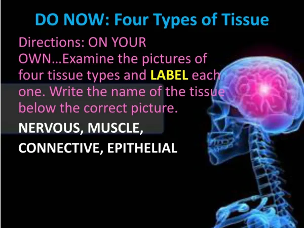
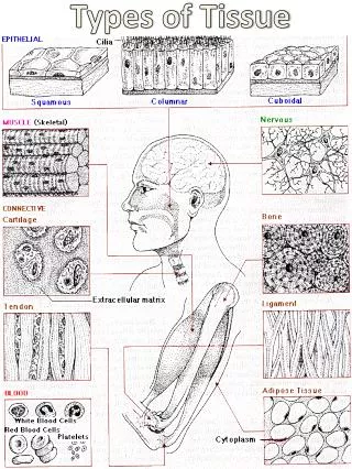
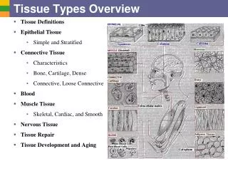
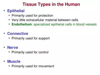
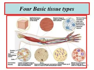
Body Tissues • Tissues • Groups of cells with similar structure and function • Four primary types… • Epithelial tissue (epithelium) • Connective tissue • Muscle tissue • Nervous tissue
Epithelium Characteristics • Cells fit closely together often sheet-like • The apical surface = free surface • The lower surface of the epithelium rests on a basement membrane • Avascular (no blood supply) • Regenerate easily if well nourished So the function and locations of these tissues would be…
Epithelium Characteristics Figure 3.17a
Epithelium Characteristics • Cells fit closely together and often form sheets • The apical surface is the free surface of the tissue • The lower surface of the epithelium rests on a basement membrane • Avascular (no blood supply) • Regenerate easily if well nourished So the function and locations of these tissues would be?
1. Epithelial Tissues • Functions • Protection • Absorption • Filtration • Secretion • Locations • Body coverings • Body linings • Glandular tissue
Classification of Epithelia • Number of cell layers • Simple—one layer • Stratified—more than one layer • Shape of cells • Squamous • flattened • Cuboidal • cube-shaped • Columnar • column-like Figure 3.17a
Simple Epithelia • Simple squamous • Single layer of flat cells • Usually forms membranes • Lines body cavities • Lines lungs and capillaries
Simple Epithelia • Simple cuboidal • Single layer of cube-like cells • Common in glands and their ducts • Forms walls of kidney tubules • Covers the ovaries
Simple Epithelia • Simple columnar • Single layer of tall cells • Often includes mucus-producing goblet cells • Lines digestive tract
Simple Epithelia • Pseudostratified columnar • Single layer, but some cells are shorter than others • Often looks like a double layer of cells • May function in absorption or secretion
Select 1 of the following types of epithelia and be able to describe its appearance, location in the body, & function • Simple squamous • Simple cuboidal • Simple Columnar • Pseudostratified columnar
Stratified Epithelia • Stratified squamous • Cells at the apical surface are flattened • Found as a protective covering where friction is common • Locations • Skin • Mouth • Esophagus
Stratified Epithelia • Stratified cuboidal—two layers of cuboidal cells • Stratified columnar—surface cells are columnar, cells underneath vary in size and shape • Stratified cuboidal and columnar • Rare in human body • Found mainly in ducts of large glands
Stratified Epithelia • Transitional epithelium • Shape of cells depends upon the amount of stretching • Lines organs of the urinary system
Glandular Epithelium • Gland • One or more cells responsible for secreting a particular product • Two major gland types • Endocrine gland • Secretions diffuse into blood vessels • All secretions are hormones • Exocrine gland • Secretions empty through ducts epithelial surface • Include sweat and oil glands
Cell Sketches! • To get used to seeing the tissue types you will be drawing a total of 6 sketches, 2 of which will be examples of epithelial tissue. • Find an example of an epithelial tissue under the microscope, critically look at it, then sketch what you see • Grading is based on • color (2 for accurate realistic, 1 realistic but the color is a little off, 0 it’s not colored or does not look realistic at all) • Determination of the approximate size (2 if within range, 0 not) • Resemblance (4 pts if the sketch looks just like the specimen proper shape, proper size @ mag., attn to detail) • Name (1 pt for recording the sketched tissue type) • Magnification (1 pt for recording the total magnification at which the specimen was viewed)
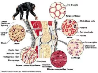
Connective Tissue • Found everywhere in the body • Includes the most abundant and widely distributed tissues • Functions • Binds body tissues together • Supports the body • Provides protection
Connective Tissue Characteristics • Variations in blood supply • Some tissue types are well vascularized • Some • have a poor blood supply = ligaments & tendons • are avascular = cartilage • Extracellular matrix • Non-living material that surrounds living cells • Made of: • Ground substance = water mainly with adhesion proteins & polysaccharides • Fibers (type and amount vary)
Connective Tissue Types • Bone (osseous tissue) • Composed of • Bone cells in lacunae (cavities) • Hard matrix of calcium salts • Large numbers of collagen fibers • Used to protect and support the body
Connective Tissue Types • Hyaline cartilage • Most common type of cartilage • Composed of • Abundant collagen fibers • Rubbery matrix • Locations • Larynx • Attach ribs to breast bone • Entire fetal skeleton
Connective Tissue Types • Elastic cartilage • Provides elasticity • Location • Supports the external ear • Tip of nose • Fibrocartilage • Highly compressible • Location • Forms cushion-like discs between vertebrae
Connective Tissue Types- Dense • Dense connective tissue (fibrous tissue) • Form strong rope-like structures • Main matrix element =collagen fibers • Locations • Tendons—attach skeletal muscle to bone • Ligaments—attach bone to bone at joints • Dermis—lower layers of the skin
Connective Tissue Types- Loose • Areolartissue • Most widely distributed connective tissue • Soft, pliable tissue like “cobwebs” • Functions as a packing tissue • Contains all fiber types • Can soak up excess fluid (causes edema)
Connective Tissue Types- Loose • Adipose tissue • Matrix is an areolar tissue in which fat globules predominate • Many cells contain large lipid deposits • Functions • Insulates the body • Protects some organs • Serves as a site of fuel storage
Connective Tissue Types- Loose • Reticular connective tissue • Delicate network of interwoven fibers • Forms stroma (internal supporting network) of lymphoid organs • Lymph nodes • Spleen • Bone marrow
Connective Tissue Types • Blood • Fluid matrix = blood plasma • Fibers are visible during clotting only • Transports materials
Cell Sketches! • To get used to seeing the tissue types you will be drawing a total of 6 sketches, 2 of which will be examples of epithelial tissue. • Find an example of an epithelial tissue under the microscope, critically look at it, then sketch what you see • Grading is based on • color (2 for accurate realistic, 1 realistic but the color is a little off, 0 it’s not colored or does not look realistic at all) • Determination of the approximate size (2 if within range, 0 not) • Resemblance (4 pts if the sketch looks just like the specimen proper shape, proper size @ mag., attn to detail) • Name (1 pt for recording the sketched tissue type) • Magnification (1 pt for recording the total magnification at which the specimen was viewed)
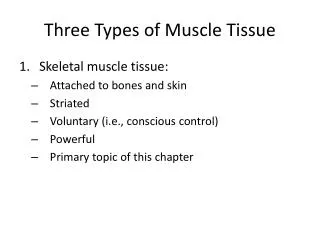
Muscle Tissue • Function is to produce movement • Three types • Skeletal muscle • Cardiac muscle • Smooth muscle
Muscle Tissue Types • Skeletal muscle • Under voluntary control • Contracts to pull on bones or skin • Characteristics of skeletal muscle cells • Striated • Multinucleate (more than one nucleus) • Long, cylindrical
Muscle Tissue Types • Cardiac muscle • Under involuntary control • Found only in the heart • Characteristics of cardiac muscle cells • Cells are attached intercalated disks • Striated • One nucleus per cell
Muscle Tissue Types • Smooth muscle • Under involuntary muscle • Found in walls of hollow organs such as stomach, uterus, and blood vessels • Characteristics of smooth muscle cells • No visible striations • One nucleus per cell • Spindle-shaped cells
Nervous Tissue • Composed of neurons and nerve support cells • Function is to send impulses to other areas of the body • Irritability—responds to a stimulus (electrical) • Conductivity—ability to carry an electrical current
Cell Sketches! • To get used to seeing the tissue types you will be drawing a total of 6 sketches, 2 of which will be examples of epithelial tissue. • Find an example of an epithelial tissue under the microscope, critically look at it, then sketch what you see • Grading is based on • color (2 for accurate realistic, 1 realistic but the color is a little off, 0 it’s not colored or does not look realistic at all) • Determination of the approximate size (2 if within range, 0 not) • Resemblance (4 pts if the sketch looks just like the specimen proper shape, proper size @ mag., attn to detail) • Name (1 pt for recording the sketched tissue type) • Magnification (1 pt for recording the total magnification at which the specimen was viewed)
Tissue Repair (Wound Healing) • Regeneration • Replacement of destroyed tissue by the same kind of cells • Fibrosis • Repair by dense (fibrous) connective tissue (scar tissue) • Determination of method • Type of tissue damaged • Severity of the injury
Events in Tissue Repair • Capillaries become very permeable • Introduce clotting proteins • A clot walls off the injured area • Formation of granulation tissue • Growth of new capillaries • Rebuild collagen fibers • Regeneration of surface epithelium • Scab detaches
Regeneration of Tissues • Tissues that regenerate easily • Epithelial tissue (skin and mucous membranes) • Fibrous connective tissues and bone • Tissues that regenerate poorly • Skeletal muscle • Tissues that are replaced largely with scar tissue • Cardiac muscle • Nervous tissue within the brain and spinal cord