Download
1 / 54
570 likes | 992 Views
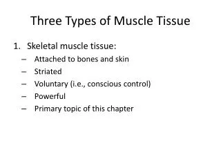
Three Types of Muscle Tissue. 1. Smooth muscle tissue 2. Skeletal muscle tissue 3. Cardiac muscle tissue. 3 Types of Muscle Tissue. Skeletal muscle attaches to bone, skin or fascia striated with light & dark bands visible with scope voluntary control of contraction & relaxation.

E N D
Three Types of Muscle Tissue 1. Smooth muscle tissue 2. Skeletal muscle tissue 3. Cardiac muscle tissue
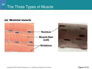
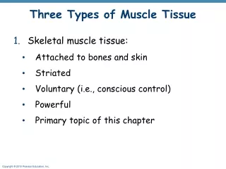
3 Types of Muscle Tissue • Skeletal muscle • attaches to bone, skin or fascia • striated with light & dark bands visible with scope • voluntary control of contraction & relaxation
3 Types of Muscle Tissue • Cardiac muscle • striated in appearance • involuntary control • autorhythmic because of built in pacemaker
3 Types of Muscle Tissue • Smooth muscle • attached to hair follicles in skin • in walls of hollow organs -- blood vessels & GI • nonstriated in appearance • involuntary
Functions of Muscle Tissue • Producing body movements • Stabilizing body positions • Regulating organ volumes • bands of smooth muscle called sphincters • Movement of substances within the body • blood, lymph, urine, air, food and fluids, sperm • Producing heat • involuntary contractions of skeletal muscle (shivering)
Properties of Muscle Tissue • Excitability • respond to chemicals released from nerve cells • Conductivity • ability to propagate electrical signals over membrane • Contractility • ability to shorten and generate force • Extensibility • ability to be stretched without damaging the tissue • Elasticity • ability to return to original shape after being stretched
Skeletal Muscle -- Connective Tissue • Superficial fascia is loose connective tissue & fat underlying the skin • Deep fascia = dense irregular connective tissue around muscle • Connective tissue components of the muscle include • epimysium = surrounds the whole muscle • perimysium = surrounds bundles (fascicles) of 10-100 muscle cells • endomysium = separates individual muscle cells • All these connective tissue layers extend beyond the muscle belly to form the tendon
Nerve and Blood Supply • Each skeletal muscle is supplied by a nerve, artery and two veins. • Each motor neuron supplies multiple muscle cells (neuromuscular junction) • Each muscle cell is supplied by one motor neuron terminal branch and is in contact with one or two capillaries. • nerve fibers & capillaries are found in the endomysium between individual cells
Fusion of Myoblasts into Muscle Fibers • Every mature muscle cell developed from 100 myoblasts that fuse together in the fetus. (multinucleated) • Mature muscle cells can not divide • Muscle growth is a result of cellular enlargement & not cell division • Satellite cells retain the ability to regenerate new cells.
Muscle Fiber or Myofibers • Muscle cells are long, cylindrical & multinucleated • Sarcolemma = muscle cell membrane • Sarcoplasm filled with tiny threads called myofibrils & myoglobin (red-colored, oxygen-binding protein)
Transverse Tubules • T (transverse) tubules are invaginations of the sarcolemma into the center of the cell • filled with extracellular fluid • carry muscle action potentials down into cell • Mitochondria lie in rows throughout the cell • near the muscle proteins that use ATP during contraction
Myofibrils & Myofilaments • Muscle fibers are filled with threads called myofibrils separated by SR (sarcoplasmic reticulum) • Myofilaments (thick & thin filaments) are the contractile proteins of muscle
Sarcoplasmic Reticulum (SR) • System of tubular sacs similar to smooth ER in nonmuscle cells • Stores Ca+2 in a relaxed muscle • Release of Ca+2 triggers muscle contraction
Atrophy and Hypertrophy • Atrophy • wasting away of muscles • caused by disuse (disuse atrophy) or severing of the nerve supply (denervation atrophy) • the transition to connective tissue can not be reversed • Hypertrophy • increase in the diameter of muscle fibers • resulting from very forceful, repetitive muscular activity and an increase in myofibrils, SR & mitochondria
Filaments and the Sarcomere • Thick and thin filaments overlap each other in a pattern that creates striations (light I bands and dark A bands) • The I band region contains only thin filaments. • They are arranged in compartments called sarcomeres, separated by Z discs. • In the overlap region, six thin filaments surround each thick filament
Thick & Thin Myofilaments • Supporting proteins (M line, titin and Z disc help anchor the thick and thin filaments in place)
Overlap of Thick & Thin Myofilaments within a Myofibril Dark(A) & light(I) bands visible with an electron microscope
Exercise-Induced Muscle Damage • Intense exercise can cause muscle damage • electron micrographs reveal torn sarcolemmas, damaged myofibrils an disrupted Z discs • increased blood levels of myoglobin & creatine phosphate found only inside muscle cells • Delayed onset muscle soreness • 12 to 48 Hours after strenuous exercise • stiffness, tenderness and swelling due to microscopic cell damage
The Proteins of Muscle • Myofibrils are built of 3 kinds of protein • contractile proteins • myosin and actin • regulatory proteins which turn contraction on & off • troponin and tropomyosin • structural proteins which provide proper alignment, elasticity and extensibility • titin, myomesin, nebulin and dystrophin
The Proteins of Muscle -- Myosin • Thick filaments are composed of myosin • each molecule resembles two golf clubs twisted together • myosin heads (cross bridges) extend toward the thin filaments • Held in place by the M line proteins.
The Proteins of Muscle -- Actin • Thin filaments are made of actin, troponin, & tropomyosin • The myosin-binding site on each actin molecule is covered by tropomyosin in relaxed muscle • The thin filaments are held in place by Z lines. From one Z line to the next is a sarcomere.
Sliding Filament Mechanism Of Contraction • Myosin cross bridgespull on thin filaments • Thin filaments slide inward • Z Discs come toward each other • Sarcomeres shorten.The muscle fiber shortens. The muscle shortens • Notice :Thick & thin filaments do not change in length
How Does Contraction Begin? • Nerve impulse reaches an axon terminal & synaptic vesicles release acetylcholine (ACh) • ACh diffuses to receptors on the sarcolemma & Na+ channels open and Na+ rushes into the cell • A muscle action potential spreads over sarcolemma and down into the transverse tubules • SR releases Ca+2 into the sarcoplasm • Ca+2 binds to troponin & causes troponin-tropomyosin complex to move & reveal myosin binding sites on actin--the contraction cycle begins
Excitation - Contraction Coupling • All the steps that occur from the muscle action potential reaching the T tubule to contraction of the muscle fiber.
Contraction Cycle • Repeating sequence of events that cause the thick & thin filaments to move past each other. • 4 steps to contraction cycle • ATP hydrolysis • attachment of myosin to actin to form crossbridges • power stroke • detachment of myosin from actin • Cycle keeps repeating as long as there is ATP available & high Ca+2 level near thin filament
Steps in the Contraction Cycle • Notice how the myosin head attaches and pulls on the thin filament with the energy released from ATP
ATP and Myosin • Myosin heads are activated by ATP • Activated heads attach to actin & pull (power stroke) • ADP is released. (ATP released P & ADP & energy) • Thin filaments slide past the thick filaments • ATP binds to myosin head & detaches it from actin • All of these steps repeat over and over • if ATP is available & • Ca+ level near the troponin-tropomyosin complex is high
Overview: From Start to Finish • Nerve ending • Neurotransmittor • Muscle membrane • Stored Ca+2 • ATP • Muscle proteins
Relaxation • Acetylcholinesterase (AChE) breaks down ACh within the synaptic cleft • Muscle action potential ceases • Ca+2 release channels close • Active transport pumps Ca2+ back into storage in the sarcoplasmic reticulum • Calcium-binding protein (calsequestrin) helps hold Ca+2 in SR (Ca+2 concentration 10,000 times higher than in cytosol) • Tropomyosin-troponin complex recovers binding site on the actin
Rigor Mortis • Rigor mortis is a state of muscular rigidity that begins 3-4 hours after death and lasts about 24 hours • After death, Ca+2 ions leak out of the SR and allow myosin heads to bind to actin • Since ATP synthesis has ceased, crossbridges cannot detach from actin until proteolytic enzymes begin to digest the decomposing cells.
Structures of NMJ Region • Synaptic end bulbs are swellings of axon terminals • End bulbs contain synaptic vesicles filled with acetylcholine (ACh) • Motor end plate membrane contains 30 million ACh receptors.
Events Occurring After a Nerve Signal • Arrival of nerve impulse at nerve terminal causes release of ACh from synaptic vesicles • ACh binds to receptors on muscle motor end plate opening the gated ion channels so that Na+ can rush into the muscle cell • Inside of muscle cell becomes more positive, triggering a muscle action potential that travels over the cell and down the T tubules • The release of Ca+2 from the SR is triggered and the muscle cell will shorten & generate force • Acetylcholinesterase breaks down the ACh attached to the receptors on the motor end plate so the muscle action potential will cease and the muscle cell will relax.
Pharmacology of the NMJ • Botulinum toxin blocks release of neurotransmitter at the NMJ so muscle contraction can not occur • bacteria found in improperly canned food • death occurs from paralysis of the diaphragm • Curare (plant poison from poison arrows) • causes muscle paralysis by blocking the ACh receptors • used to relax muscle during surgery • Neostigmine (anticholinesterase agent) • blocks removal of ACh from receptors so strengthens weak muscle contractions of myasthenia gravis • also an antidote for curare after surgery is finished
Muscle MetabolismProduction of ATP in Muscle Fibers • Muscle uses ATP at a great rate when active • Sarcoplasmic ATP only lasts for few seconds • 3 sources of ATP production within muscle • creatine phosphate • anaerobic cellular respiration • anaerobic cellular respiration
Anaerobic Cellular Respiration • ATP produced from glucose breakdown into pyruvic acid during glycolysis • if no O2 present • pyruvic converted to lactic acid which diffuses into the blood • Glycolysis can continue anaerobically to provide ATP for 30 to 40 seconds of maximal activity (200 meter race)
Aerobic Cellular Respiration • ATP for any activity lasting over 30 seconds • if sufficient oxygen is available, pyruvic acid enters the mitochondria to generate ATP, water and heat • fatty acids and amino acids can also be used by the mitochondria • Provides 90% of ATP energy if activity lasts more than 10 minutes
Muscle Fatigue • Inability to contract after prolonged activity • central fatigue is feeling of tiredness and a desire to stop (protective mechanism) • depletion of creatine phosphate • decline of Ca+2 within the sarcoplasm • Factors that contribute to muscle fatigue • insufficient oxygen or glycogen • buildup of lactic acid and ADP • insufficient release of acetylcholine from motor neurons
Oxygen Consumption after Exercise • Muscle tissue has two sources of oxygen. • diffuses in from the blood • released by myoglobin inside muscle fibers • Aerobic system requires O2 to produce ATP needed for prolonged activity • increased breathing effort during exercise • Recovery oxygen uptake • elevated oxygen use after exercise (oxygen debt) • lactic acid is converted back to pyruvic acid • elevated body temperature means all reactions faster
Muscle Tone • Involuntary contraction of a small number of motor units (alternately active and inactive in a constantly shifting pattern) • keeps muscles firm even though relaxed • does not produce movement • Essential for maintaining posture (head upright) • Important in maintaining blood pressure • tone of smooth muscles in walls of blood vessels
Classification of Muscle Fibers • Slow oxidative (slow-twitch) • red in color (lots of mitochondria, myoglobin & blood vessels) • prolonged, sustained contractions for maintaining posture • Fast oxidative-glycolytic (fast-twitch A) • red in color (lots of mitochondria, myoglobin & blood vessels) • split ATP at very fast rate; used for walking and sprinting • Fast glycolytic (fast-twitch B) • white in color (few mitochondria & BV, low myoglobin) • anaerobic movements for short duration; used for weight-lifting
Fiber Types within a Whole Muscle • Most muscles contain a mixture of all three fiber types • Proportions vary with the usual action of the muscle • neck, back and leg muscles have a higher proportion of postural, slow oxidative fibers • shoulder and arm muscles have a higher proportion of fast glycolytic fibers • All fibers of any one motor unit are same. • Different fibers are recruited as needed.
Anabolic Steroids • Similar to testosterone • Increases muscle size, strength, and endurance • Many very serious side effects • liver cancer • kidney damage • heart disease • mood swings • facial hair & voice deepening in females • atrophy of testicles & baldness in males
Anatomy of Cardiac Muscle • Striated , short, quadrangular-shaped, branching fibers • Single centrally located nucleus • Cells connected by intercalated discs with gap junctions • Same arrangement of thick & thin filaments as skeletal
Cardiac versus Skeletal Muscle • More sarcoplasm and mitochondria • Larger transverse tubules located at Z discs, rather than at A-l band junctions • Less well-developed SR • Limited intracellular Ca+2 reserves • more Ca+2 enters cell from extracellular fluid during contraction • Prolonged delivery of Ca+2 to sarcoplasm, produces a contraction that last 10 -15 times longer than in skeletal muscle
Physiology of Cardiac Muscle • Autorhythmic cells • contract without stimulation • Contracts 75 times per min & needs lots O2 • Larger mitochondria generate ATP aerobically • Sustained contraction possible due to slow Ca+2 delivery • Ca+2 channels to the extracellular fluid stay open
Two Types of Smooth Muscle • Visceral (single-unit) • in the walls of hollow viscera & small BV • autorhythmic • gap junctions cause fibers to contract in unison • Multiunit • individual fibers with own motor neuron ending • found in large arteries, large airways, arrector pili muscles,iris & ciliary body
Physiology of Smooth Muscle • Contraction starts slowly & lasts longer • no transverse tubules & very little SR • Ca+2 must flows in from outside • Calmodulin replaces troponin • Ca+2 binds to calmodulin turning on an enzyme (myosin light chain kinase) that phosphorylates the myosin head so that contraction can occur • enzyme works slowly, slowing contraction
Smooth Muscle Tone • Ca+2 moves slowly out of the cell • delaying relaxation and providing for state of continued partial contraction • sustained long-term • Useful for maintaining blood pressure or a steady pressure on the contents of GI tract
Regeneration of Muscle • Skeletal muscle fibers cannot divide after 1st year • growth is enlargement of existing cells • repair • satellite cells & bone marrow produce some new cells • if not enough numbers---fibrosis occurs most often • Cardiac muscle fibers cannot divide or regenerate • all healing is done by fibrosis (scar formation) • Smooth muscle fibers (regeneration is possible) • cells can grow in size (hypertrophy) • some cells (uterus) can divide (hyperplasia) • new fibers can form from stem cells in BV walls