Download
1 / 3
30 likes | 50 Views
To know more about Visit our website. Visit:- https://www.uscultrasound.com/

E N D
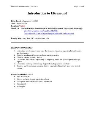
Structure of the Human Body (2021/2022) Amy Kule, MD Introduction to Ultrasound Date: Tuesday, September 28, 2020 Time: Asynchronous Location:Virtual Watch Medical Student Introduction to Bedside Ultrasound Physics and Knobology: https://www.youtube.com/watch?v=RDqWK- DzPus&list=PL3Zty8IubWpexaYymphlUdPrnlsVBR-Oi&index=8 Faculty Info: Amy Kule, MD – akule@lumc.edu LEARNING OBJECTIVES Understand how to maneuver around the ultrasound machine regarding button location and functionality Describe tranducer differences and appropriate selection Describe various scanning modes Understand function and adjustmeny of frequency, depth and gain to optimize image capture Understand scanning terminology: hyperechoic, hypoechoic, anechoic Describe and demonstrate scanning planes: longitudinal (sagittal), transverse (axial), coronal HANDS-ON OBJECTIVES Turn machine on Choose and activate appropriate transducer Place probe and indicator in correct orientation Adjust depth Adjust gain
Structure of the Human Body (2021/2022) Amy Kule, MD INTRODUCTION TO ULTRASOUND Why Ultrasound? –Portable, fast, inexpensive, non-invasive, no ionizing radiation Limitations of Ultrasound –Depends on skill of user (“operator-dependent”) and patient body habitus. Ultrasound does not pass well through bone, and is scattered by air ALARA (As Low As Reasonably Achievable) – A philosophy of radiation use based on keeping exposure as low as possible. How Does Ultrasound Work? –Piezoelectric Effect Image Credit: Eric Shappell Loyola ‘13 When electricity is applied to piezoelectric crystals, the vibrations produce sound waves that travel outward. The waves hit objects and bounce back to the piezoelectric crystals, and the mechanical energy produced from the vibration of the crystal is converted into electrical energy. The ultrasound machine calculates the time it takes for sound waves to travel back to the probe and produces a gray-scale image on the screen. Knobology: -Find ‘ON’ button -Depth -Gain Probes: - Linear, Curvilinear, Phased Array, Endocavitary - High frequency probes vs Low frequency probes (Resolution, Penetration)
Structure of the Human Body (2021/2022) Amy Kule, MD Basic Motions: -Sweep -Slide -Fan -Rotate -Rock (Heel-Toe) Ultrasound Convention: -Probe marker (Bump) to the patient’s right or head Scanning Planes: -Longitudinal (Sagittal) -Transverse (Axial) -Coronal Basic Terms: -Echogenic – Structure or medium capable of reflecting or transmitting ultrasound waves, creating a brighter image -Anechoic – Image appears dark (black) Example: Fluid-filled structures such as bladder, blood vessel -Hyperechoic – Ultrasound Image Appears Brighter Than Surroundings (White) Example: Bone -Hypoechoic - Ultrasound image appears darker than surrounding structures -Isoechoic – Ultrasound image appears similar in brightness to surrounding structures Additional Resources: 1.Ultrasound FundamentalsVideo by Eric Shappell (SSOM ’13) http://www.youtube.com/watch?v=RXvvQYONlmE 2.Machine Orientation Video by Eric Shappell (SSOM ’13) https://www.youtube.com/watch?v=ReATA_9o_gE 3.Ultrasound Basics USC Ultrasound Learning Modules http://uscm.med.sc.edu/mods/1B/player.html