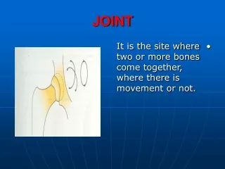
Joint Features
Joint Features . Solid joints: structural, little movement, synarthroses (fibrous); symphyses (cartilage) Synovial joints: cavity, movement Capsule, ligaments, muscles Synovial lining, phagocytic or fibroblast-like cells, hyaluronic acid lubricant

Joint Features
E N D
Presentation Transcript
Joint Features • Solid joints: structural, little movement, • synarthroses (fibrous); symphyses (cartilage) • Synovial joints: cavity, movement • Capsule, ligaments, muscles • Synovial lining, phagocytic or fibroblast-like cells, hyaluronic acid lubricant • hyaline cartilage shock absorber, wear resistant • chondrocytes synthesise and digest matrix of type 2 collagen, proteoglycans
Osteoarthritisprogressive degeneration of articular cartilage • Usually age-related and oligoarticular; vertebrae, hips, knees, feet and hands (DIP, PIP) • May be secondary to obesity, trauma, or joint disease (haemophilia) • Pain, morning stiffness, crepitus, decreased movement, root compression, nerve pressure effectes • Cartilage degenerates, chondrocytes cluster, increased water, decreased proteoglycans, fibrillation, fragmentation, joint mice, bone exposed, fractures, cysts, eburnation, osteophytes (Heberden’s nodes), reactive synovium, fibrous pannus • Chondrocytes produce Il-1, TNF alpha, Il-6, prostaglandins
Rheumatoid Arthritischronic systemic inflammatory disorder • 1% of population, majority (x5) women, peak 20-40 years • Involves many sites: skin (rheumatoid nodules), blood vessels (vasculitis), heart, lungs • Principally involves joints: hands, feet; later wrists, elbows, ankles, knees • Onset: gradual systemic or acute arthritis, swollen, hot, painful and stiff joints
Rheumatoid Arthritis • Pathogenesis • Genetic susceptiblity (HLA Dr4, common motif of antigen binding site of DR molecule) • Antigen: microbial? Epstein Barr, mycoplasma, borrelia • Autoimmunity: to collagen or cartilage proteoglycan, driven by CD4 cells • Joint damage: • activated cells cytokine loop, • B cell activation, IgM autoantibodies to IgG (rheumatoid factor) • immune complexes • Effect: • inflamed synovium, oedema, thickened hyperplastic papillae form • Increased vascularity, lymphocytes, plasma cells, fibrin • Bone eroded, subchondral cysts, osteopenia, • pannus (granulation tissue obliterating synovium); ankylosis
8 other forms of arthritis • Juvenile rheumatoid arthritis: large joints, oligo involvement, RF negative, ANA+ • Ankylosing spondylitis, HLA B27, males, axial, sacroiliac joints • Reiter syndrome: Arthritis, urethritis, conjunctivitis, autoimmune reaction following infection of GI or urethra • Psoriatic arthritis: 5% of sporiasis, like RA • Infectious: suppurative, tuberculous • Lyme arthritis: borrelia burgdorferei, late in disease, large joints • Gout: uric acid crystals, acute arthritis, chronic tophaceous arthritis, tophi • Pseudogout: calcium pyrophosphate, knees
Vitamin D • Role: bone mineralisation, maintaining serum calcium and phosphate levels • Source; skin, 7 dehydrocholesterol (sun) vitamin D3: diet ergosterol altered to vitamin D2 • Synthesis: travels on binding protein • to liver (hydroxylase) produces 25 (OH)D • to kidney (hydroxylase) produces 1.25 (OH)D • Effects • GI absorbs calcium and phosphate • Bone: mobilises calcium phosphate (with PTH) • Kidney: absorbs calcium, loses phosphate (with PTH)
Rickets and osteomalacia • Vitamin D deficiency: • 1) diet (sunlight), • 2 malabsorption, • 3 metabolism: liver or kidney disease (cyto P450, renal hydroxylase deficiency), • 4 end organ resistance • Phosphate deficiency (aluminium bound, failed renal tubular absorption • Normal growing cartilage - becomes calcified - stops growing - endochondral ossification • Children (rickets): growing cartilage not calcified, abnormal growth and poor ossification, abnormal bone in skull, ribs, vertebrae, lower limbs • Adult (osteomalacia): growth finished, excess osteoid, osteopenia, fractures
Osteoporosis • Peak bone mass; effect of vitamin D, diet, exercise, hormones • Normal bone remodeling: bone loss less than 1% a year • Senile osteoporosis: with age, osteoblasts less effective (diet and exercise influence) increased slow bone loss mainly in cortex • Post menopausal osteoporosis: decreased oestrogen allows increase in cytokines (Il-1, Il-6 and TNF alpha) which mobilizes osteoclasts; reactive osteoblast mobilisation, high turnover bone loss, main effects on cancellous bone • Result: (microfracture) vertebrae—pain, height loss, deformity; pelvis, hips—fractures (fatal secondary illnesses; pneumonia, pulmonary embolism)
HyperparathyroidismRenal osteodystropy • Primary or secondary hyperparathyroidism: increased PTH signals osteoblasts which signal osteoclasts to resorb bone • Effects: • Subperiosteal resorption (hand X-ray) • Dissecting osteitis: resorption, fibrosis, microfractures of cancellous bone • Brown tumour (osteitis fibrosa cystica, Von Recklinghausen) haemorrhage, haemosiderin, inflammatory repair, cystic change • Renal osteodystrophy : • Renal failure causes defective 1,25 (OH)D, loss of phosphate and retention of H (acidosis) • These induce changes of hyperparathyroidism, osteomalacia, osteoporosis, osteosclerosis and decreased growth. The bone loss may be high, low or mixed turnover • Excess aluminium and amyloid may add to the effect
Bone Features • Bone profile: joint - cartilage - epiphysis - plate - metaphysis - diaphysis (shaft) • Bone section: periosteum - cortical bone - cancellous bone - medulla • Periosteum: produces reactive bone if stimulated (causes Codman’s triangle, sunburst, or onion skin patterns on X-ray) • Osteoblasts, contain alkaline phosphate, become osteocytes, produce osteoid (like collagen) which becomes mineralised as bone (fibres are at first irregular - woven - later become parallel - lamellar • Osteoclast: multinucleated cell of monocyte-histiocyte lineage, contains acid phosphate, lyses bone. • Growth: at metaphysis, site of most tumours • Long bone growth: metaphyses near knee (lower femur, upper tibia) remote from elbow (upper humerus) • Effects of age: closure of epiphyses
Bone tumours • Presentation: pain, tenderness, swelling, deformity, pathological fracture • 1) secondary tumours: 70% of cancer involves bone; spreads to bone by blood or vertebral lymphatics • Deposits are usually: multiple, in axial skeleton, in the red marrow, lytic, may sclerotic (prostate, breast) • (role of vessels, prostaglandins, osteoclast activing factor) • Effect: hypercalcemia, leukoerythroblastic anaemia • 2) marrow tumours: Leukaemia, lymphoma, myeloma • 3) primary bone tumours: benign or malignant
Osteosarcoma • Primary (70%): 10-25 years, males x 2, some familial or follow retinoblastoma • Secondary (30%): paget’s disease, enchondromatosis, chronic osteomyelitis, fibrous dysplasia, bone infarcts or fractures, radiation (radium or external), alkalating agents • Aetiology: ? Virus, immunological, genetic, related to bone mass • Usual findings: in medulla of long bones at metaphysis, 70% about the knee as bulky mass • Codman’s triangle on x-ray • 25% have metastases (usually lung) at diagnosis, mean survival 3 years • Variants: may be cortical, periosteal, parosteal, extraskeletal • Microscopy: osteoblastic (but also chondroblastic, fibroblastic, angioblastic areas)
Chondrosarcoma • Males x 2, age 50s and 60s • Primary (90%) • Secondary (10%) enchondromas etc. as for osteosarcoma • Presentation: lobulated mass with spotty calcification; pelvic bone (50%) or other central sites • Correlation of clinical, x-ray and histopathology essential (malignant central lesion histologically similar to a benign peripheral lesion) • Microscopy: most are low grade (grade 1) 90% 5-year survival; grade 3 have poor survival (40% 5-year survival)
Ewing’s Sarcoma • Presentation: Pain, tenderness, swelling, systemic symptoms (may simulate osteomyelitis), age 5-20, males x 2, involves pelvis or long bones, 25% have metastases (to lung, brain, other bones) at diagnosis • Begins in medulla of metaphysis or of diaphysis but spreads to periosteum producing Codman’s triangle and onion layering effect on x-ray • Microscopy: small round cells with cytoplasmic glycogen (PAS+) • Cell of origin unknown: ?mesenchymal, related to peripheral neuroectodermal tumour (PNET) which has neural feature; both have t(11-22), produce a fusion gene EWS-FLI1, stain with antibodies to CD99 • Survival: has improved from 10% to 75% with modern chemotherapy
Giant cell tumour • Microscopy: neoplastic stromal cells with prominent giant cells • Exclude other lesions which may have giant cells including brown tumour of hyperparathyroidism, gingival epulis, fibrous cortical defect, aneurysmal bone cyst, chondroblastoma. Carcinoma and soft tissue sarcoma may sometimes have prominent giant cells but rarely cause diagnostic difficulties • Occur in epiphysis after closure (age 15-55 years), 50% about knee, more frequent in females • Most are benign, one-third to one-half recur, 10% are aggressive or malignant but such behaviour can not reliably be predicted histologically
Paget’s disease • Bone disease with osteoclastic (early), mixed , and osteosclerotic (late) stages • May be monostotic or polyostotic; usually involves axial skeleton • Involves 1-3% of the older population, males (4:3) more frequently • Prodominantly a disease of people of N European stock, ? Viral cause, viral type particles in osteoclasts • Usually asymptomatic (identified incidentally on x-ray or by raised alkaline phosphatase). Causes excess production of collagen products (hydroxyproline) • May cause pain, deformity, nerve pressure, haemodynamic shunting of blood, pathological fracture, sarcoma • Therapy: calcitonin, diphosphonates, mithramycin, fluorides
Fibrous dysplasia • A developmental defect showing impaired bone formation with disorganised osseous and fibrous tissue which fractures and causes deformities • Usually monostotic, ribs, skull, jaw, femur (in teens and 20s) • In 25% polyostotic (usually children) • Occasionally (3%) with pigmentation, precocious puberty (females) and endocrinopathy (hyperthyroidism, hyperparathyroidism, hyperpituitarism, diabetes) as Allbright’s syndrome
Other cartilage tumours Osteochondroma • Disordered growth from epiphysis to form abnormal metaphyseal spur • Mainly a sporadic disorder of long bones in adolescence • Occasionally multiple as an autosomal dominant disorder • Rarely sarcoma may supervene Enchondroma • Sporadic single lesions, may be asymptomatic or cause deformity • Multiple (enchondromatosis) (Ollier’s disease) as a non-hereditiary disease involving hand and feet in childhood • Multiple with multiple cavernous haemangiomas as a familial disease (Maffucci’s disease) Rare lesions: Chondromyxoid fibroma, Chondroblastoma
Other bone tumours Osteoma • Rare, usually involves face of skull • Gardner’s syndrome: osteomas with epidermal cysts, teeth disorders, desmoid tumours, and colonic adenomatous polyps Osteoid osteoma • Tiny (3-10 mm) vascular osteoblastic lesion (nidus), regarded as a benign neoplasm, surrounded by a large reactive osteosclerotic rim • Larger, histologically similar lesion in vertebrae and lower limbs is painless (osteoclastoma) • Causes severe night pain relieved by aspirin, cured by excision • Age 5-25, males x 3 • Site: metaphysis of long bones especially of lower limb
Cysts • Simple: solitary fluid-filled lesions in metaphysis near epiphyseal plate. Two-thirds in proximal long bones, males x 3, childhood and adolescence • Aneurysmal bone cyst: blood-filled spaces separated by fibrous septae distend bone and may extend into surrounding tissue (may be secondary to degeneration of another lesion). Involve vertebrae and flat bones (10-20 years; M=F) • Fibrous cortical defect (non-ossifying fibroma): Defect of epiphyseal plate growth extending into the metaphysis, usually about the knee, in children