Joint
Joint. By Dr. Ajay Kumar Professor School of Physical Education DAVV Indore. Introduction.

Joint
E N D
Presentation Transcript
Joint By Dr. Ajay Kumar Professor School of Physical Education DAVV Indore
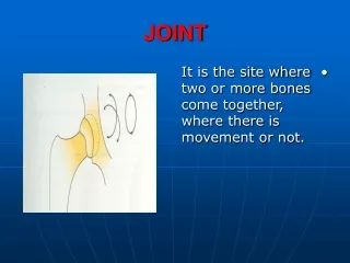
Introduction • We have already learnt that the human skeleton consists of more than 200 bones. The individual bones are attached in such a way that a large variety of co-ordinated movements are made possible in different parts of the body. These movements are made possible by skeletal muscles, the fact that the bones act as levers, cartilage which reduces fricton and ligaments which prevent dislocation and the presence of movable joints. The site or place where 2 or more bones of the skeleton are attached to each other is called a joint or place of articulation.
Joint Definition • A joint can be defined as follows: • A joint or place of articulation is formed where 2 or more bones come inclose contact in the body and are attached to each other by ligaments or cartilage.
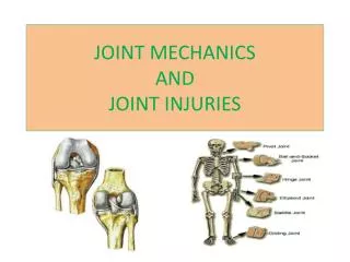
Types of Joints • Joints can be classified according to the degree and type of movement they allow. The following types of joints can be recognized:
Fibrous (or Immovable) Joints. • These joints are firmly held together by a thin layer of strong connectice tissue. There is no movement between the bones such as the sutures of the skull and the teeth in their sockets.
Cartilagenous Joints • Cartilagenous joints are joints where the articular surfaces of the bones forming the joints are attached to each other by means of white fibrocartilaginous discs and ligaments which allow only a limited degree of movement. Examples are the cartilaginous between the vertebrae, the cartilage in the symphysis which binds the pubic bones together at the front of the pelvic girdle and the cartilage in the joint between the sacrum and the hip bone.
Synovial Joints • These are freely movable joints. Most of the joints in the body are of the synovial type. The following are the main characteristics of a synovial joint: • The ends of the bones are covered with a layer of smooth hyaline cartilage, called articular cartilage in the joint regions. This reduces fricton at the point. • The joint is completely enclosed by a bag-like capsular ligament which holds the joint together and helps to contain the synovial fluid.
Synovial Joints (cont) • The capsular ligament is lined with a synovial membrane. This membrane secretes synovial fluid into the synovial cavity and acts as a seal, waterproofing the joint. The synovial fluid lubricates the joint. • In addition to the capsule, the bones are also attached and held together by strong, tough ligaments made of dense connective tissue. These ligaments prevent dislocation during normal movement. • The articulating surfaces of adjacent bones are reciprocally shaped.
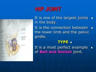
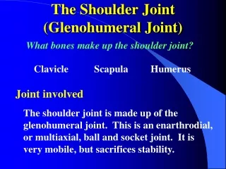
Ball-and-socket joint • Consists of a bone with a globular or slightly egg-shaped head that articulates with the cup-shaped cavity of another bone. Such a joint allows a wider range of motion than does any other kind, permitting movements in all planes, as well as rotational movement around a central axis. The hip and shoulder contain joints of this type.
Condyloid joint • The ovoid condyle of one bone fits into the elliptical cavity of another bone, as in the joints between the metacarpals (bones of the palm) and phalanges (bones of the fingers and toes). This type of joint permits a variety of movements in different planes; rotational movement, however, is not possible.
Gliding joints • The articulating surfaces are nearly flat or slightly curved. These joints allow sliding or back-and-forth motion and twisting movements. Most of the joints within the wrist and ankle, as well as those between the articular processes of adjacent vertebrae, belong to this group. The sacroiliac joints and the joints formed by ribs 2 though 7 connecting with the sternum are also gliding joints.
Hinge joint • The convex surface of one bone fits into the concave surface of another, as in the elbow and the joints of the phalanges. Such a joint resembles the hinge of a door in that it permits movement in one plane only.
Pivot Joint • The cylindrical surface of one bone rotates within a ring formed of bone and fibrous tissue of a ligament. Movement at such a joint is limited to rotation around a central axis. The joint between the proximal ends of the radius and the ulna, where the head of the radius rotates in a ring formed by the radial notch of the ulna and a ligament (annular ligament), is of this type. Similarly, a pivot joint functions in the neck as the heard turns from side to side. In this case, the ring formed by a ligament (transverse ligament) and the anterior arch of the atlas rotates around the dens of the axis.
Saddle joint • Forms between bones whose articulating surfaces have both concave and convex regions. The surface of one bone fits the complementary surface of the other. This physical relationship permits a variety of movements, mainly in two planes, as in the case of the joint between the carpal (trapezium) and the metacarpal of the thumb.