Download

1 / 47
470 likes | 1.31k Views
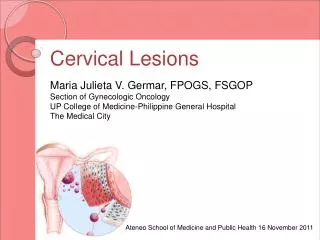
Vulvar and Vaginal lesions. Dr.F Behnamfar MD. Introduction. Most usful means of generating differential diagnosis is by morphological findings rather than symptomatology

E N D
Vulvar and Vaginal lesions Dr.FBehnamfar MD
Introduction Most usful means of generating differential diagnosis is by morphological findings rather than symptomatology Vulvar biopsy should be performed if the lesion is clinically suspicious or does not resolve after standard therapy
Vulvar Symptoms • Most often,primary vaginitis and secondary vulvitis • A number of skin conditions on other areas of the body
Neoplasia • Vulvar intraepithelial neoplasia a precancerous lesion that may progress to invasive cancer • Most are raised multifocal white (may be red or pink) and/or verrucous lesions • Cancer presents with unifocal vulvar plaque,ulcer or mass • Lichen scerosus and erosive lichen planus predispose to cancer
Genital warts • Caused by human papillomavirus • Flat,filliform or verrucous,or giant • Flesh colored or pigmented • Biopsy is indicated if there is rapid growth,increased pigmentation,ulceration,pigmentation,fixation or poor response to therapy • Treatment : trichloroacetic acid, podophyllum,Cryo,laser • Not curative ,merely speed clinical resolution
White patch • Lichen sclerosus,well demarcated white finely wrinkled and atrophic patches • Vulvar itching and typical findings • Potent topical corticosteriod ointment • Close follow up for risk of malignancy
Other vulvar conditions • folliculitis • Fox.fordiyce disease • Acanthosis nigricans • Extramammary pagets disease,intraepithelial adenocarcinoma
Herpes simplex • Scabis
Vulvar cysts, tumors and masses • Condylomata accuminata • duct cysts,Skenes duct cysts • Vulvar Ulcers: Behcet disease,lichen planus
Vaginal Conditions • Retained foreign body • Ulceration • Malignancy
Vulvar Cancer • 3870 new cases 2005 • 870 deaths • Approximately 5% of Gynecologic Cancers American Cancer Society. Cancer Facts & Figures. 2004. Atlanta, GA; 2005
Vulvar Cancer • 85% Squamous Cell Carcinoma • 5% Melanoma • 2% Sarcoma • 8% Others
Vulvar Cancer • Biphasic Distribution ,two distinct etiologies: • Age 70 • type, unifocal, • in areas adjacent to lichen sclerosus or squamous hyperplasia (Chronic inflammatory conditions) • 20% in patients UNDER 40 and appears to be increasing, • multifocal, • basaloid or warty types, • HPV related,smoking and VIN
Vulvar Cancer • Paget’s Disease of Vulva • 10% will be invasive • 4-8% association with underlying Adenocarcinoma of the vulva
Symptoms • Most patients are treated for “other” conditions • 12 month or greater time from symptoms to diagnosis
Symptoms • Pruritus • Mass • Pain • Bleeding • Ulceration • Dysuria • Discharge • Groin Mass
Symptoms • May look like: • Raised • Erythematous • Ulcerated • Condylomatous • Nodular
Vulvar Cancer • IF IT LOOKS ABNORMAL ON THE VULVA • BIOPSY! • BIOPSY! • BIOPSY!
Tumor Spread • Very Specific nodal spread pattern • Direct Spread • Hematogenous
Staging • Based on TNM Surgical Staging • Tumor size • Node Status • Metastatic Disease
Staging • Stage I T1 N0 M0 • Tumor ≤ 2cm • IA ≤1 mm depth of stromal Invasion • IB 1 mm or more depth of invasion
Staging • Stage II T2 N0 M0 • Tumor >2 cm • Confined to Vulva or Perineum
Staging • Stage III • T3 N0 M0 • T3 N1 M0 • T1 N1 M0 • T2 N1 M0 • Tumor any size involving lower urethra, vagina, anus OR unilateral positive nodes
Staging • Stage IVA • T1 N2 M0 • T2 N2 M0 • T3 N2 M0 • T4 N any M0 • Tumor invading upper urethra, bladder, rectum, pelvic bone or bilateral nodes
Staging • Stage IVB • Any T Any N M1 • Any distal mets including pelvic nodes
Treatment • Primarily Surgical • Wide Local Excision • Radical Excision • Radical Vulvectomy with Inguinal Node Dissection • Unilateral • Bilateral • Possible Node Mapping, still investigational
Treatment • Local advanced may be treated with Radiation plus Chemosensitizer • Positive Nodal Status • 1 or 2 microscopic nodes < 5mm can be observed • 3 or more or >5mm post op radiation
New advances in treatment • Individualization of treatment,vulvar conservation for unifocal tumors • Elimination of routine pelvic lymphadenectomy • Omission of groin dissection for T1 tumors (<1mm stromal invasion) • Separate incisions improve wound healing
Treatment • Special Tumor • Verrucous Carcinoma • Indolent tumor with local disease, rare mets UNLESS given radiation, becomes Highly malignant and aggressive • Excision or Vulvectomy ONLY
Vulva 5 year survival • Stage I 90 • Stage II 77 • Stage III 51 • Stage IV 18 Hacker and Berek, Practical Gynecologic Oncology 4th Edition, 2005
Recurrence • Local Recurrence in Vulva • Reexcision or radiation and good prognosis if not in original site of tumor • Poor prognosis if in original site
Recurrence • Distal or Metastatic • Very poor prognosis, active agents include Cisplatin, mitomycin C, bleomycin, methotrexate and cyclophosphamide
Melanoma • 5% of Vulvar Cancers • Not UV related • Commonly periclitoral or labia minora
Melanoma • Microstaged by one of 3 criteria • Clark’s Level • Chung’s Level • Breslow
Melanoma Treatment • Wide local or Wide Radical excision with bilateral groin dissection • Interferon Alpha 2-b
Vaginal Carcinoma • 2140 new cases projected 2005 • 810 deaths projected 2005 • Represents 2-3% of Pelvic Cancers American Cancer Society. Cancer Facts & Figures. 2004. Atlanta, GA; 2005
Vaginal Cancer • 84% of cancers in vaginal area are secondary • Cervical • Uterine • Colorectal • Ovary • Vagina Fu YS, Pathology of the Uterine Cervix, Vagina and Vulva, 2nd ed. 2002
Vaginal Carcinoma • Squamous Cell 80-85% • Clear Cell 10% • Sarcoma 3-4% • Melanoma 2-3%
Clear Cell Carcinoma • Associated with DES Exposure In Utero • DES used as anti abortifcant from 1949-1971 • 500+ cases confirmed by DES Registry • Usually occurred late teens
Vaginal Cancer Etiology • Mimics Cervical Carcinoma • HPV 16 and 18
Staging • Stage I Confined to Vaginal Wall • Stage II Subvaginal tissue but not to pelvic sidewall • Stage III Extended to pelvic sidewall • Stage IVA Bowel or Bladder • Stage IVB Distant mets
Treatment • Surgery with Radical Hysterectomy and pelvic lymph dissection in selected stage I tumors high in Vagina • All others treated with radiation with chemosensitization
5 year Survival • Stage I 70% • Stage II 51% • Stage III 33% • Stage IV 17%