Black and Brown Lesions
440 likes | 942 Views
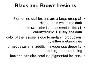
Black and Brown Lesions. Pigmented oral lesions are a large group of disorders in which the dark or brown color is the essential clinical characteristic. Usually, the dark color of the lesions is due to melanin production by either melanocytes

Black and Brown Lesions
E N D
Presentation Transcript
Black and Brown Lesions • Pigmented oral lesions are a large group of disorders in which the dark • or brown color is the essential clinical characteristic. Usually, the dark • color of the lesions is due to melanin production by either melanocytes • or nevus cells. In addition, exogenous deposits and pigment-producing • bacteria can also produce pigmented lesions.
Benign disorders, deposits ,benign and malignant neoplasms, and systemic diseases are included in the group of pigmented lesions : O Normal pigmentation O Amalgam tattoo O Heavy-metal deposition O Drug-induced pigmentation O Smoker’s melanosis O Black hairy tongue O Ephelis O Lentigo O Lentigo maligna O Pigmented nevi O Nevus of Ota O Melanoma O Addison disease O Peutz–Jeghers syndrome
Normal Pigmentation • Definition and etiology Increased melanin production and deposition • in the oral mucosa may often be a physiological finding, particularly in • dark-skinned individuals. • Clinical features This type of pigmentation is persistent and symmetrical, • and clinically presents as asymptomatic black or brown areas of • varying size. The gingiva are most commonly affected, followed by the • buccal mucosa, palate, and lips The pigmentation is more • prominent in areas of pressure or friction, and usually becomes more • intense with increasing age. • Laboratory tests Histopathological examination. • Differential diagnosis Addison disease, smoker’s melanosis, drug-induced • pigmentation, pigmented nevi, melanoma, amalgam tattoo. • Treatment No treatment is required.
Amalgam Tattoo • Definition Amalgam deposition (tattoo) is a common oral disorder. • Etiology Implantation of dental amalgam into the oral mucosa. • Clinical features The condition presents as a well-defined irregular or diffuse flat area, with a bluish-black discoloration of varying size The most common sites of involvement are the gingiva, alveolar • mucosa, and buccal mucosa. The diagnosis is usually made at the clinical level • Laboratory tests Histopathological examination and radiographs. • Differential diagnosis Pigmented nevi, lentigo, freckles, melanoma, • normal pigmentation, other metal tattoo. • Treatment No treatment is required.
Heavy-Metal Deposition • Definition and etiology Heavy-metal deposition is a rare oral condition caused by ingestion or exposure to bismuth, lead, silver, mercury, and other heavy metals. • Clinical features Clinically, the most common pattern (bismuth, lead) is a bluish line along the marginal gingiva, or similar spots within the gingival papillae .Rarely, diffuse bluish-black discoloration may be seen (silver). The diagnosis is based on the history and the clinical features. • Differential diagnosis Normal pigmentation, amalgam tattoo. • Treatment No treatment is required for oral lesions.
Drug-Induced Pigmentation • Definition Drug-induced oral pigmentation is a relatively common • condition, caused by increased melanin production or drug metabolite • deposition. • Etiology Antimalarials, tranquilizers, minocycline, azidothymidine, ketoconazole, phenolphthalein, and others are the most common drugs • that induce pigmentation. • Clinical features The clinical picture varies, and the condition may • appear as irregular brown or black macules or plaques, or diffuse melanosis • The buccal mucosa, tongue, palate, and gingiva are the • most commonly affected sites. The diagnosis is made on the basis of the • history and clinical criteria. • Differential diagnosis Normal pigmentation, Addison disease, Peutz– • Jeghers syndrome. • Treatment No treatment is required.
Smoker’s Melanosis • Definition Smoker’s melanosis, or smoking-associated melanosis, is a benign abnormal melanin pigmentation of the oral mucosa. • Etiology Tobacco smoke that stimulates melanocytes. • Clinical features Clinically, it appears as multiple brown pigmented • areas, usually located on the anterior labial gingiva of the mandible • Pigmentation of the buccal mucosa and palate has been associated • with pipe smoking. The intensity of pigmentation is related to • time and dose. Women are more commonly affected. • Differential diagnosis Normal pigmentation, drug-induced pigmentation, pigmented nevi, melanoma, Addison disease. • Treatment No treatment is required. Cessation of smoking is usually • associated with a return of normal mucosal pigmentation.
Black Hairy Tongue • Hairy tongue may occasionally appear black as a result of the growth of pigment-producing bacteria that colonize the elongated • Filiform papillae .In addition, the black color may also be due to staining from food and tobacco. The diagnosis is made on the basis of • clinical criteria.
Ephelis • Definition: Ephelides, or freckles, are discrete brown macules, commonly • seen on sun-exposed skin and rarely in the mouth. • Etiology Unknown. They are due to increased melanin production. • Clinical features Clinically, the lesions appear as solitary and well demarcated asymptomatic round brown macules, less than 5 mm in • diameter .The vermilion border of the lower lip is the most • common site of development. • Laboratory tests Histopathological examination. • Differential diagnosis Lentigo, pigmented nevi, melanoma, drug-associated pigmentation, Peutz–Jeghers syndrome, Albright syndrome • Treatment No treatment is required, except for aesthetic or diagnostic • considerations. • .
Lentigo • Definition Lentigo is a rare oral disorder of pigmentation. • Etiology Increased number of epidermal melanocytes. • Clinical features The condition presents as small round flat spots, • brown or dark brown in color, usually less than 0.5 cm in diameter • It is a rare lesion intraorally. • Laboratory tests Histopathological examination. • Differential diagnosis Ephelis, pigmented nevi, melanoma, Peutz– • Jeghers syndrome. • Treatment No treatment is required
Lentigo Maligna • Definition Lentigo maligna, or Hutchinson’s freckle, is a premalignant • lesion of melanocytes that probably represents in-situ melanoma. • Etiology Unknown. • Clinical features Lentigo maligna is very rare intraorally. Clinically, it • appears as a slowly expanding black or brown plaque, with irregular • borders . In 5–15 years, it ultimately progresses into invasive • melanoma. The lips, buccal mucosa, palate, and floor of the mouth are • the common sites affected. • Laboratory tests Histopathological examination. • Differential diagnosis Melanoma, pigmented nevi, amalgam tattoo. • Treatment Surgical excision, radiotherapy. • .
Pigmented Nevi • Definition Pigmented cellular nevi are benign malformations of melanocytes • and “nevus cells,” common in the skin and rare in the oral • mucosa. • Etiology Developmental. Melanocytes and nevus cells of neural crest • origin. • Clinical features Based on histological criteria, oral pigmented nevi • are classified into four types: intramucosal, junctional, compound, and • blue. Clinically, the lesion appears as an asymptomatic, well-demarcated, • flat or slightly elevated, brown, black, or blue spot or plaque . The • lesion is usually solitary, with a diameter of less than 1 cm. The palate, • gingiva, buccal mucosa, and lips are the sites of predilection. • Laboratory tests Histopathological examination. • Differential diagnosis Ephelis, lentigo, melanoma, amalgam tattoo. • Treatment Usually, no treatment is required. Conservative surgical • excision is carried out in some cases
Nevus of Ota • Definition Nevus of Ota, or oculodermal melanocytosis, is a hamartomatous disorder of the melanocytes that predominantly involves the skin of the face and eyes, and mucous membranes. Characteristically, the lesions follow the distribution of the first and second branches of the trigeminal nerve • Etiology Developmental. Hyper pigmentation is due to melanin-producing melanocytes in the dermis that have failed to reach the epidermis or epithelium during fetal life
Clinical features The skin lesions present as multiple mottled black or brown macules varying in size from 1 mm to several millimeters . The oral lesion presents as asymptomatic blue or blue-black dots or patches that usually involve the palate and buccal mucosa . Hyperpigmentation of ipsilateral sclera is a common sign, while involvement of the cornea, iris, fundus, oculi, and retina is rare. Other sites such as nasal mucosa, pharynx, and tympanum may be less commonly affected. The disorder usually appears in early childhood before the age of 1 year and around puberty. About 70–80% of the cases are female. Malignant transformation of nevus of Ota is very rare. The diagnosis is mainly based on the history and the clinical features. Laboratory tests Histopathological examination. Treatment Laser and camouflage of face lesions
Melanoma • Definition Melanoma is a malignant neoplasm originating either de • novo from melanocytes, or from a benign melanocytic lesion. • Etiology Unknown. Ultraviolet radiation is an important causative • factor for skin melanoma. • Clinical features Primary oral melanoma is uncommon, representing • 0.5–1.5% of melanomas. Clinically, it presents as a black or brown macule, • plaque, or nodule that may be ulcerated . The lesions • are usually characterized by an irregular margin and a tendency to • spread. Based on clinical and histopathological criteria, oral melanoma • is classified into three forms: lentigo maligna melanoma (best prognosis), • superficial spreading melanoma (good prognosis), and nodular melanoma • (poor prognosis). The palate, upper gingiva, and alveolar mucosa are • most commonly affected. • Laboratory tests Histopathological examination. • Differential diagnosis Pigmented nevi, ephelis, lentigo, lentigo maligna, • amalgam tattoo, pyogenic granuloma, Kaposi sarcoma. • Treatment Surgical excision, radiotherapy, chemotherapy.
Multiple nodular malignant melanomas of the alveolar mucosa of the maxilla
Addison Disease • Definition Addison disease is a relatively uncommon insufficiency of • adrenal corticosteroid hormones. • Etiology Adrenal cortex destruction, usually caused by autoimmunity, • infections, tumors, amyloidosis. • Clinical features The oral manifestations are common and early, and • present as diffuse or patchy dark brown pigmentation, due to melanin • production . The buccal mucosa, palate, lips, and gingiva are • the most common sites of involvement. • Laboratory tests Measurement of plasma adrenocorticotropic hormone • (ACTH) and serum cortisol levels. • Differential diagnosis Normal pigmentation, drug-induced pigmentation, • Peutz–Jeghers syndrome. • Treatment Steroid replacement.
Peutz–Jeghers Syndrome • Definition Peutz–Jeghers syndrome is a rare genetically transmitted • disorder, characterized by mucocutaneous pigmentation and intestinal • polyposis. • Etiology Inherited as an autosomal dominant trait. • Clinical features The oral manifestations are the most important diagnostic • findings, and consist of oval or round, brown or black macules or • spots, 1–10 mm in diameter . The perioral skin, lips, buccal • mucosa, and tongue are the most common sites affected. The skin lesions • consist of numerous, usually perioral, dark spots . Intestinal • polyps (hamartomas) are constant findings, usually in the jejunum and • ileum. • Laboratory tests Histopathological examination, radiography of the • gastrointestinal tract. • Differential diagnosis Ephelides, lentigo, normal pigmentation, Addison • disease. • Treatment Supportive; surgical intervention in some cases.
Peutz–Jeghers syndrome: multiple pigmented spots on the buccal mucosa.
Peutz–Jeghers syndrome: multiple round spots on the lower lip.
Peutz-Jeghers syndrome, multiple pigmented spots on the skin.
Another classification • ▼ BLUE/PURPLE VASCULAR LESIONS • Hemangioma • Varix • Angiosarcoma • Kaposi’s Sarcoma • Hereditary Hemorrhagic Telangiectasia • ▼ BROWN MELANOTIC LESIONS • Ephelis and Oral Melanotic Macule • Nevocellular Nevus and Blue Nevus • Malignant Melanoma • Drug-Induced Melanosis • Physiologic Pigmentation • Café au Lait Pigmentation • Smoker’s Melanosis • Pigmented Lichen Planus • Endocrinopathic Pigmentation • HIV Oral Melanosis • Peutz-Jeghers Syndrome
▼ BROWN HEME-ASSOCIATED LESIONS Ecchymosis Petechia Hemochromatosis ▼ GRAY/BLACK PIGMENTATIONS Amalgam Tattoo Graphite Tattoo Hairy Tongue Pigmentation Related to Heavy-Metal Ingestion