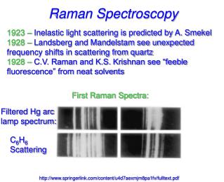
Raman Spectroscopy
Raman Spectroscopy. Komal Choudhary Lecturer School of Biotechnology DAVV Indore. When radiation passes through a transparent medium, the species present scatter a fraction of the beam in all directions.

Raman Spectroscopy
E N D
Presentation Transcript
Raman Spectroscopy Komal Choudhary Lecturer School of Biotechnology DAVV Indore
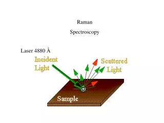
When radiation passes through a transparent medium, the species present scatter a fraction of the beam in all directions. • In 1928, the Indian physicist C. V. Raman discovered that the visible wavelength of a small fraction of the radiation scattered by certain molecules differs from that of the incident beam and furthermore that the shifts in wavelength depend upon the chemical structure of the molecules responsible for the scattering. • Raman spectroscopy deals with the inelastic scattering of light and not with its absorption.
Electric polarizability(α)- it is defined as the ratio of the induced dipole movement P of an atom to the electric field E that produce this dipole movement. • P= αE • For asymmetric molecules, absorptions will give rise to both types and virtually the same information could be obtained from either. However for symmetrical molecules(CO2,C2H2) having a centre of symmetry, the fundamental frequencies that appear in the Raman do not appear in the IR and vice-versa(“Mutual exclusion rule” ). these two methods are truly complementary. • For Exp.- the stretching vibration of homonuclear diatomic molecule(such as H2, O2, N2) which are inactive in IR(because the dipole movement is zero) are observed in raman spectra.
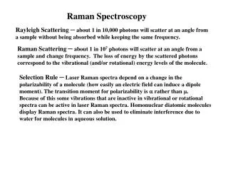
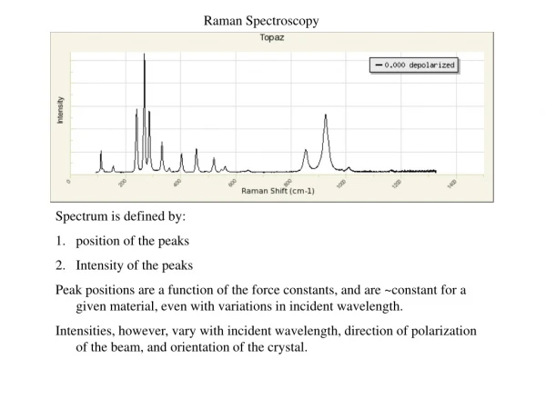
Rayleigh observed that if a substance is irradiated with monochromatic light the scattered light is observed in a direction at right angle to the incident light. • Rayleigh scattering- The frequency of scattered light is same as the frequency of incident radiation . • Stokes scattering- The frequency of scattered light is less than the frequency of incident radiation . • Antistokes scattering- The frequency of scattered light is greater than the frequency of incident radiation .
Energy Scheme for Photon Scattering Virtual State hn0+hnm hn0 Energy hn0 hn0 hn0 hn0-hnm E0+hnm E0 Rayleigh Scattering (elastic) Stokes Scattering Anti-Stokes Scattering IR Absorption Raman (inelastic) The Raman effect comprises a very small fraction, about 1 in 107 of the incident photons.
Stokes scattering: energy lost by photon: • — (( — )) Photon in Photon out No vibration Vibration • Anti-Stokes scattering: energy gained by photon: (( — )) — Photon in Photon out Vibration No vibration
But dominant process is elastic scattering: • Rayleigh scattering • — — Photon in Photon out No vibration No vibration If incident photon energy E; vibration energy v, then in terms of energy, photon out has energy: E-v Stokes scattering E+vanti-Stokes scattering E Rayleigh scattering (elastic scattering)
Light source-Mercury arc, four low pressure mercury discharge tube (increase intensity), laser. • Sample holder- glass, Quartz sample used for analysis in Raman spectroscopy may be liquid(water used for solvent), solid, or gases(sample holder generally bigger in size) . • Collection optics- lens and Notch filter(NF) lens directs the scattered radiation upon the slit of the spectrograph and the Raman lines are obtained on the photographic plate . NF is used to suppress the Rayleigh scattering. • Spectrograph- it should posses the following characteristics: • It should have large gathering power. • Special prisms of high resolving power should be employed. • A short focus camera should be employed.
Application • In inorganic chemistry structure of CO2 and structure of N2O. structure of mercurous salts. • In physical chemistry The amorphous state of a substance give rise to broad and diffused bands while crystalline state gives fine sharp line. The intensity of Raman lines enable us to determine the number and nature of ion produced by electrolytic dissociation, therefore, we can decide whether the dissociation is complete or partial. • In organic chemistry it has been observed that each functional group will have its own characteristic frequency
In biochemical research Raman Spectroscopy mainly used for intermediated sized molecule such as drugs, metabolic intermediates and substrates for exp: identification of substances such as penicillin and its derivatives, small peptides and environmental pollutants. It is an ideal rapid method for measuring certain contaminants in foodstuffs. • Use in study of photosynthesis and respiration in plants, particularly for CO2 metabolism.