Raman Spectroscopy
Raman Spectroscopy. Rayleigh Scattering ─ about 1 in 10,000 photons will scatter at an angle from a sample without being absorbed while keeping the same frequency. .

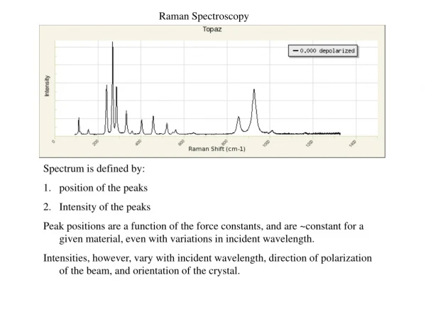
Raman Spectroscopy
E N D
Presentation Transcript
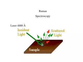
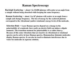
Raman Spectroscopy Rayleigh Scattering ─ about 1 in 10,000 photons will scatter at an angle from a sample without being absorbed while keeping the same frequency. Raman Scattering ─ about 1 in 107 photons will scatter at an angle from a sample and change frequency. The loss of energy by the scattered photons correspond to the vibrational (and/or rotational) energy levels of the molecule. Selection Rule ─ Laser Raman spectra depend on a change in the polarizability of a molecule (how easily an electric field can induce a dipole moment). The transition moment for polarizability is a rather than m. Because of this some vibrations that are inactive in vibrational or rotational spectra can be active in laser Raman spectra. Homonuclear diatomic molecules display Raman spectra. It can also be used to eliminate interference due to water for molecules in aqueous solution.
Electronic Spectra - atoms Hydrogen - n ℓ mℓ. none Dℓ = ±1 Dmℓ = 0, ±1 Since ℓ and mℓ don’t affect energies, the spectrum is fairly simple. ^ M = Ym*mYndt Multi-electron quantum numbers L S J 0, ±1 0 0, ±1 For 2 electrons: L = ℓ1 + ℓ2 → |ℓ1 - ℓ2| = 0, 1, or 2 S = s1 + s2 → |s1 - s2| = 0, 1 J = L + S Fig. 15.7 pg 534 Term Symbols:2S+1LJ. Table 15.1 pg 529
Possible Term Symbols Term Symbols:2S+1LJ. Table 15.1 pg 529 s12S p1 & p5 2P p2 & p4 1S, 1D, 3P p32P, 2D, 2S d1 & d9 2D d2 & d8 1S, 1D, 1G, 3P, 3F d3 & d7 2P, 2D, 2F, 2G, 2H, 4P, 4F L = 0 1 2 3 4 5 6 S P D F G H I
Electronic Spectra - molecules Multi-electron quantum numbersS Ω Term Symbols:2S+1Ω = 0 1 2 3 S P D F 0, ±1 0 0, ±1
p*p transitions – These typically result from C=C double bonds. The energies are about 7eV (about 180 nm) for unconjugated systems. The more conjugated the system the closer the energy levels are together, and the higher the transition wavelength. They can occur in the near UV (e.g. Trp residues in proteins) to the visible range (many types of dyes, e.gCoomassie blue used to stain proteins). The absorption of rhodopsin in the retina is an example of a p-p* transition in the visible range. Spartan (AM1) HOMO → LUMO note that molecular modeling eVl energies are poor and the actual Cyclohexene 10.82 115 l values are much higher Benzene 10.21 122 (180)but the general trend is accurate. Napthalene 8.45 147 (275)
p*n transitions – Lone pair O or N electrons in C=O or C=N bonds can cause p*n transitions. For example carbonyls absorb in the near UV between 270-290 nm. However, these transitions are much less intense than p*p transitions. This is why the UV spectra of proteins are dominated by aromatic groups rather than carbonyls, although the latter do contribute.
H3N+ - CH – COO- CH2 N .. FF = # photons emitted = _____kf______ # photons absorbed (kf + knr + kic) The Chronology of florescence (see fig. 1) Absorbance A + hnaA*t ~ 10-15 s Vibrational de-excitation A* A* + heatt ~ 10-12 s Fluorescence emission A* A + hnet ~ 10-8 s rate = kf non-radiative decay A* A + heatt ~ ??? rate = knr
LASER Light Amplification by Stimulated Emission of Radiation Metastable excited State ─ has long t Population Inversion ─ A majority of molecules exist in mes rather than gs Pumping ─ gs → es → mes via high intensity, short duration l pulse Stimulated Emission ─ mes → gs using l pulse = to laser output Coherence ─ l output photons are all in same phase (waves are synchronized) spatial and temporal phase
Four Level Laser I A stimulating l and laser output pumping l A' X
Three Level Laser I A pumping l stimulating l and laser output X