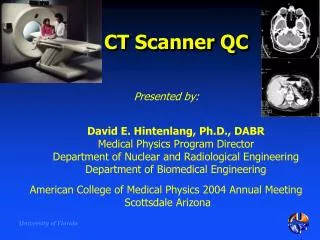
CT Scanner QC
CT Scanner QC. Presented by:. David E. Hintenlang, Ph.D., DABR Medical Physics Program Director Department of Nuclear and Radiological Engineering Department of Biomedical Engineering. American College of Medical Physics 2004 Annual Meeting Scottsdale Arizona.

CT Scanner QC
E N D
Presentation Transcript
CT Scanner QC Presented by: David E. Hintenlang, Ph.D., DABR Medical Physics Program Director Department of Nuclear and Radiological Engineering Department of Biomedical Engineering American College of Medical Physics 2004 Annual Meeting Scottsdale Arizona
CT Quality Control - Motivations • Appropriate diagnostic benefit vs risk • Image Quality • Noise • Contrast • Spatial resolution • Radiation Risk • Dosimetry indices (ie. CTDI)
Presentation Overview • Introduction & Background • General categories of QC tests • Specific QC tests and procedures • QC Program recommendations & comparisons • Acceptance tests • “Annual” QC tests • “Daily” QC tests • Conclusions & References
Background • Expanded CT capabilities over last decade • Helical and Multislice CT • Decreases scan times - Changes in patient dose • Increased utilization of CT • Complex systems, high utilization, large contribution to patient dose demands a continuous and robust QA program
QC Objectives • Complexity of system offers opportunities for many individual QC tests • Level of testing balanced against QC Objectives • QC Categories: • Acceptance Testing • Annual Testing • Daily QC • ACR Accreditation
Categories of QC Tests • Safety • Mechanical accuracy • Imaging Performance • Display System
General QC Tests : Safety • Electrical • Mechanical • Laser • Radiation
General QC Tests: Mechanical Accuracy • Ensure that aligned patient anatomy is imaged • Alignment light accuracy • Alignment of table and gantry • Table and gantry positioning • Slice localization from scout images
General QC Tests: Image Quality • Noise and Field Uniformity • CT Number Linearity • Low Contrast Detectability • Spatial Resolution • Display and Hard Copy Image Quality
General QC Tests: Dosimetry • CTDI • Head & Body • Patient Dosimetry
Alignment Laser Accuracy • Internal & External scan plane lights • Verify correct distance between internal & external scan plane lights • Wrap film around centered phantom • Make pin hole marks along illuminated lines
Scan 1 slice @ 1mm thickness • Repeat for external alignment laser • Measure distance between line of pierced holes and center of exposed slice • Misalignment should be less than 2 mm
Table - Gantry Alignment • To ensure that longitudinal axis of table is aligned with isocenter • Measure to locate & mark central axis of table at: • head end • middle • foot end • Hang plumb bob from center of gantry - to table top • Measure distances between plumb bob and central axis of table • Repeat for various gantry angles • Table should be aligned within +/- 5 mm
Table Incrementation Accuracy • Evaluate accuracy/ reproducibility of longitudinal table motion • Tape ruler along table edge • Tape pointer on frame around mid-point of ruler • Load table • Move table 300-500 mm in one direction & return • Repeat for a total of 3-times in each direction • Average & Standard Deviation should be less than 3 mm
Table Incrementation Accuracy & Collimation (axial scan) Evaluate accuracy & reproducibility of longitudinal table motion & width of x-ray beam • Wrap film around centered head phantom • Scan 5 slices for several combinations of slice thickness and gap. • Measure slice thickness and gaps from film • Mark table frame at one end of the phantom • Return to original table position and note any difference from original position Interslice gap should have < 0.5 mm discrepancy Slice thickness should be accurate to within 1 mm
Table Incrementation, scanned volume, helical pitch accuracy Evaluate accuracy & reproducibility of longitudinal table motion & helical pitch • Wrap film around centered head phantom • Mark table frame at one end of phantom • Record indicated table position • Obtain volume scans for various slice thicknesses and pitch Measure from film: Total length scanned Spiral slice thickness Spiral gaps (pitch)
kVp Accuracy • Lock x-ray tube position • Center voltage divider using CT specific filer pack in center of x-ray beam • Check kVp’s at : 80, 100, 140 kVp Tube potential should be within +/- 5%
Half-Value Layer • Lock x-ray tube position • Set scanning parameters per manufacturer (80 kVp and mAs to obtain 200 mR) • Mount CT chamber parallel to table long axis, centered on isocenter • Vary Al thicknesses Should be at least 2.3 mm Al @ 80 kVp
Exposure Reproduceabiity & Linearity • Head phantom in head holder on table • Insert CT ion chamber in center of phantom • Perform 4 scans at 80 kVp, 100 mA, 1s • Record exposure for each • Repeat at: 200 mA, 300 mA • Repeat at : 120 kVp, 140 kVp
Radiation Profile Width Evaluates the accuracy of pre-patient collimator settings • Tape film around centered head phantom positioned flat on table with 0o gantry angle • Scan one slice at each slice thickness Lowest kVp and tube current available space about 2 cm apart • Measure width of each slice Should be within 1 mm of selected thickness
Slice Sensitivity Profile Evaluate the actual width of the imaged slice • Set standard scanning parameters • Scan phantom insert with “ramps” for each available slice thickness • For each image determine average pixel values of: a) metal ramp and b) acrylic background • Set window width < 10 HU of min width setting • Set window level at average of ramp and background values • Measure width of ramp and multiply by tanq
Image Quality Measures • Noise and Field Uniformity • CT Number Linearity • Low Contrast Detectability • Spatial Resolution • Reconstruction times • Scout view accuracy
Phantoms & Test Tools • Testing phantoms available from manufacturers & third parties • Early CT phantoms may not provide sensitivity require to test modern CT systems
Noise & Field UniformityAxial Scan Assess noise level under simulated clinical conditions Set standard scanning parameters Position a centered homogenous phantom Scan and select ROI’s Obtain mean pixel value and standard deviation for each ROI Std. Dev. Central ROI < 10 HU Max difference between peripheral and central ROI’s < +/- 5 HU
Noise & Field UniformityVolume Scan Assess noise level under simulated clinical conditions • Set standard scanning parameters • Position a centered homogenous phantom • For each voume scanned measure 3 reconstructed slices • center & both ends of volume • Scan and select ROI’s • Obtain mean pixel value and standard deviation for each ROI Std. Dev. Central ROI < 10 HU Max difference between peripheral and central ROI’s < +/- 5 HU
CT Number Linearity CT numbers should be consistent from system to system • Set standard scanning parameters • Image phantom insert with 5 rods of different materials • Determine mean pixel values of each object & background • Perform for low, mid, & high kVp settings CT number should be within 50 HU for each material
Low Contrast Detectability Evaluate scanner low contrast object discrimination • Set standard scanning parameters • Scan low-contrast detectability insert • Determine mean value of each hole & surrounding material • Contrast = difference in HU of objects & background • Determine Number of objects visible Should meet manufacturer’s specifications
Spatial Resolution Evaluate high contrast spatial resolution • Set standard scanning parameters • Scan Spatial Resolution Insert • Determine highest frequency group visually resolved for each image Should meet manufacturer specifications
Spatial Resolution MTF Derive the MTF as a measure of limiting spatial resolution • Scan spatial resolution insert • For each group of high contrast resolution patterns determine the mean value & std. deviation. • Determine & plot MTF • Repeat using bone reconstruction filters Should meet manufacturer’s specifications
Image Reconstruction Time Evaluate reconstruction times • Set parameters for typical head scan • Scan anthropomorphic head phantom • Measure time required to obtain 1 image slice • Subtract out data collection time • Repeat for anthropomorphic chest phantom and typical body slice Should meet manufacturer specifications
Scout Views Evaluate scout view production • Position anthropomorphic head phantom • Perform scout view at 120 kVp, 160 mA, 1 mm thickness • Scan length 0 500 mm • Perform scout views for 0, 45 and 90 degree gantry angles • Record time required to perform and print images • Repeat with anthropomorphic chest phantoms
Display and Hard Copy Image Quality Ensure that information in the video signal is accurately transferred to display media • Reduce room illumination to normal viewing level • Display SMPTE Pattern • Adjust brightness and contrast controls 5% and 95% patches should be visible
Dosimetry Measurement: CTDI Determine a measure of radiation dose to tissue under different scan conditions • Set standard scanning parameters (i.e. head) • Position CTDI head phantom • Scan with CT ion chamber inserted sequentially in each of 5 positions • Calculate CTDI using peripheral and central values • Repeat with CTDI body phantom • Repeat for various scanning parameters as desired
CT Equipment Quality Control Program • ACR Accreditation Program for CT • QC program established & implemented under supervision of a qualified medical physicist • Requirements • Acceptance testing upon installation • Annual medical physicist survey • Continuous Quality Control
Acceptance Testing • Series of measurements performed to verify that a CT system conforms to required specifications • Specific tests/technical specifications should be included in bid request or purchase agreement • Many more combinations of operating parameters exist than can be practically tested • Must acquire sufficient data to adequately characterize system performance
Considerations for Acceptance Tests • Determine most important performance specs to test in view of: • Clinical needs • Scanner capabilities • Devise a short list of 4-5 “standard” scanning conditions: • Standard body • Standard head • Spine • Pediatric Body • Pediatric Head • Pertinent performance tests should be performed for each standard condition to provide a performance baseline • Select a few additional scanner conditions that test performance limits
Essential Tests Localization light accuracy Table Incrementation Radiation Profile Sensitivity Profile Image Noise Field Uniformity High Contrast Resolution Display & Hard Copy Image Quality Radiation Dose Optional Tests Alignment of table to gantry Table gantry tilt mAs Linearity kVp Accuracy Scan Field Dependence (QCT) Alternative Tests Slice localization from Scout image High contrast resolution – MTF Low contrast detectability AAPM Report #39 Recommendations
Localization light accuracy Alignment of table to gantry Table gantry tilt Slice localization from Scout image Table Incrementation Slice thickness -Sensitivity Profile Image Noise Field Uniformity High Contrast Resolution Low Contrast Detectability Artifact Evaluation CT Number Accuracy & Linearity Display & Hard Copy Image Quality Radiation Dose – CTDI Patient Radiation Dose for select exams Safety Evaluation Visual inspection Audible/visual signals Posting requirements Scattered radiation measurements Other regulatory tests Annual ACR Requirements
Localization light accuracy Slice localization from Scout image Table Incrementation Slice thickness -Sensitivity Profile Image Noise Field Uniformity High Contrast Resolution Low Contrast Detectability Artifact Evaluation CT Number Accuracy & Linearity Display & Hard Copy Image Quality Radiation Dose – CTDI Dosimetry from Scout Image Safety Evaluation Visual inspection Audible/visual signals Posting requirements Scattered radiation measurements Specified by ACR but not AAPM 74 Patient Radiation Dose for select exams Alignment of table to gantry Table gantry tilt Monthly to Semi-Annual Tests per AAPM Report # 74
Continuous Quality Control • Required for all CT units • Qualified medical physicist should determine tests, frequency, and responsible individual • Facility Considerations • CT Usage • On-site radiological technologist should be identified with responsibility for conducting routine QC
Continuous Quality Control Tests Localization light accuracy Slice thickness Image Noise Field Uniformity High Contrast Resolution Low Contrast Detectability Artifact Evaluation CT Number Accuracy Display & Hard Copy Image Quality
Daily Tests- AAPM Report #74 CT Number Accuracy Image Noise Field Uniformity Artifact Evaluation All can be obtained from same CT phantom scans Sensitive to a wide variety of scanner problems
Aids to Daily QC i.e. AutoQA LiteTM1 • A software program to evaluate and monitor performance of CT scanners • Provides database tool for tracking a limited number of tests: • Image Noise • CT Number accuracy • Field Uniformity • Slice Width • Spatial Resolution 1. The Institute for Radiological Image Sciences, Inc. , MedSoft Technologies, Inc.
AutoQA Lite Overview • Provides automated analysis of CT phantom images CatPhan series ACR Accreditation phantom Toshiba InnerVision Vendor specific (GE, Siemens, and Philips) • Operates on PC with Dicom 3.0 CT image formats
Conclusions • ACR provides an excellent outline for CT QC Programs • A wide variety of tests exist • Medical Physicist has latitude to define requirements for “continuous” QC testing
CT QC References: • American College of Radiology Website • www.acr.org (links to “accreditation”) • AAPM Report No. 39, Specification and acceptance testing of computed tomography scanners, American Institute of Physics, 1993. • www.aapm.org/pubs/reports/ • AAPM Report No. 74, Quality control in diagnostic radiology, Medical Physics Publishing, 2002. • www.aapm.org/pubs/reports/ • ImPACT, CT Scanner Acceptance Testing, Version 1.02, 2001 • www.impactscan.org/acceptance.htm • The ACR CT Accreditation Program and the Medical Physicist, Maynard High, ACMP 2003 Annual Meeting • www.acmp.org/meetings.html