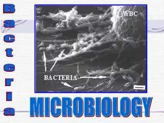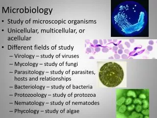
Microbiology
Microbiology. Chapter 11 Characterizing and Classifying Prokaryotes. We can characterize prokaryotes by structure. Morphology of Prokaryotic Cells Most diverse group of cellular microbes Thrive in various habitats Only a few are capable of colonizing humans and causing disease

Microbiology
E N D
Presentation Transcript
Microbiology Chapter 11 Characterizing and Classifying Prokaryotes
We can characterize prokaryotes by structure • Morphology of Prokaryotic Cells • Most diverse group of cellular microbes • Thrive in various habitats • Only a few are capable of colonizing humans and causing disease • Exist in a variety of shapes
Figure 11.1 Typical prokaryotic morphologies. “rod” “curved”
General Characteristics of Prokaryotic Organisms • Endospores • Produced by the Gram-positive bacteria Bacillus and Clostridium • Each vegetativecell transforms into one endospore • Each endospore germinates to form one vegetative cell • Defensive strategy against unfavorable conditions • Concern to: • food processors (food must be destroyed) • health care professionals (extra-hard cleaning and decontamination) • governments (public health hazard; food unable to be sold)
General Characteristics of Prokaryotic Organisms – SUMMARY SLIDE • Reproduction of Prokaryotic Cells • All reproduce asexually • Three main methods: • Binary fission (most common) • Snapping division (old cell wall does not break for daughter cells until they get big enough) • Budding
Figure 11.3 Binary fission (in gram positive bacteria). Each cell begins making its own cell wall as the septum forms The two cells physically rip themselves away from each other and their old cell wall
Figure 11.4 Snapping division, a variation of binary fission (in gram positive).
General Characteristics of Prokaryotic Organisms • Arrangements of Prokaryotic Cells • NOT the same as colony morphology! • Visible only under microscope • Seen from solid culture or gentle liquid culture • Results from two aspects of division during binary fission: • Planes by which cells divide • Must the daughter cells separate from each other to divide again
Know each of these arrangements of cocci. Single division plane; cells separate easily Or stop dividing when bound to neighbor Single division plane; cells do not separate two division planes at right angles; cells stop dividing when bound to neighbors three division planes at right angles; cells stop dividing when bound to neighbors RANDOM division planes; cells do not need to separate to keep dividing
Know each of these arrangements of bacilli. cells separate easily Single division plane (short axis); cells stop dividing when bound to neighbor Single division plane (short axis); cells do not stop dividing Single division plane; cells do not stop dividing; snapping division keeps them clustered
Modern Prokaryotic Classification • Currently based on genetic relatedness of 16S or 18S rRNA sequences • This rRNA channels the mRNA into and through the ribosome • critical function for ribosome • TIGHTLY conserved through billions of years • Three Domains of life on Earth: • Bacteria (sorted by 16S rRNA gene sequence) • Archaea (sorted by 18S rRNA gene sequence) • Eukarya (sorted by 18S rRNA gene sequence)
Figure 11.10 Prokaryotic taxonomy – SUMMARY SLIDE • Archaea • Extremophiles • thermophiles, halophiles, methanogens • Bacteria • Gram (+) bacteria – phylogenetically older group • low G+C • Clostridium, Mycoplasma, Bacillus, Listeria, Lactobacillus, Streptococcus, Enterococcus, Staphylococcus, • high G+C • Corynebacterium, Mycobacterium, Actinomycetes • Gram (-) bacteria – phylogenetically younger group • Proteobacteria • alpha-, beta-, gamma, delta-, epsilon-, zeta- • Chlamydia, Spirochetes, Bacterioides
Survey of Archaea • Generally not pathogenic • Extremophiles • Require extreme conditions to survive • Temperature, pH, and/or salinity • Prominent members are thermophiles(> 60 C) and halophiles (> 9% NaCl)
Survey of Archaea • Methanogens • Largest group of archaea • Convert carbon dioxide, hydrogen gas, and organic acids to methane gas • Convert organic wastes in pond, lake, and ocean sediments to methane • Some live in colons of animals (cows) • One of primary sources of environmental methane • Have produced ~10 trillion tons of methane that is buried in mud on ocean floor
Survey of Bacteria • Low G + C Gram-Positive Bacteria • Clostridia (Clostridium or Clostridioidessp.) • Rod-shaped, obligate anaerobes • Many form endospores spontaneously • In preparation for exposure to oxygen-rich world until reingested into an oxygen-poor environment • Important in medicine, industry, and bioenergy • C. difficile – gastroenteritis from overuse of antibiotics • C. perfringens – gas gangrene • C. tetani – tetanus • C. botulinum – botulism, botox-based therapies (inhibit muscle spasms, relax muscles for cosmetics • C. thermocellum – cellulose-based ethanol production
Survey of Bacteria • Low G + C Gram-Positive Bacteria • Mycoplasma species • Facultative or obligate anaerobes • Lack cell walls (Which antibiotics will not work???) • Smallest free-living cells – cannot resolve most cells clearly in a light microscope • Colonize mucous membranes of the respiratory (atypical pneumonia) and urinary tracts (vaginoses, pelvic inflammatory disease) • Major contaminant of mammalian cell culture
Survey of Bacteria • Low G + C Gram-Positive Bacteria • Bacillus • Many are common in soil • Form endospores – highly resistant to UV • Bacillus anthracis- anthrax • Bacillus cereus- grain-based food poisoning • Bacillus thuringiensis toxin used by farmers and gardeners as an insecticide • (cloned toxin gene used to create Bt transgenic vegatables)
Survey of Bacteria • Low G + C Gram-Positive Bacteria • Listeria species • L. monocytogenes– listeriosis through contaminated milk and meat products • Capable of reproducing under refrigeration • Can cross the placenta in pregnant women • Lactobacillus species • Grows in the body naturally but rarely causes disease • Used in the production of various foods (yogurt, kefir) • Primary genus used in probiotic formulations
Survey of Bacteria • Low G + C Gram-Positive Bacteria • Streptococcusand Enterococcus • Cause numerous diseases (mucosa and gut) • Various strains of multi-drug-resistant streptococci • MDR enterococci often precede MDR streptococci and staphylococci • Staphylococcus • Cause numerous disease (skin, mucosa) • One of the most common inhabitants of humans • Produces toxins and enzymes that contribute to disease, especially when patient or skin weakened
Survey of Bacteria • High G + C Gram-Positive Bacteria • Corynebacterium • Pleomorphic aerobes and facultative anaerobes • Most are harmless • C. diphtheriae - diphtheria • Produces metachromatic granules (phosphates) and V-shaped arrangements of cells • Mycobacterium • Aerobic rods that sometimes form filaments • Slow growth partly due to mycolic acid in its cell walls • Mycolic acid makes it resistant to antibiotics, detergents, and the immune system • M. tuberculosis – tuberculosis (consumption) • M. leprae – Hansen’s disease (leprosy)
Survey of Bacteria • High G + C Gram-Positive Bacteria • Actinomycetes • Form branching filaments resembling fungi • Important genera include Actinomyces, Streptomyces • Generally live in the soil and are not pathogenic • Nocardia can colonize human oral mucosa if inhaled or ingestde • Some Nocardia species can cause opportunistic oral or gingival disease • can progress to pneumonia, endocarditis or encephalitis with high mortality
Survey of Bacteria • Gram-Negative Proteobacteria • Largest and most diverse group of gram-neg bacteria • At least six classes of proteobacteria: • Alphaproteobacteria • Betaproteobacteria • Gammaproteobacteria • Deltaproteobacteria • Epsilonproteobacteria • Zetaproteobacteria
Survey of Bacteria • Gram-Negative Proteobacteria • Class Alphaproteobacteria • Nonpathogenic species • Nitrogen fixers and nitrifying bacteria • Grow in association with the roots of plants • Oxidation of nitrogenous compounds provides electrons • Important in the environment and agriculture
Survey of Bacteria • Gram-Negative Proteobacteria • Class Alphaproteobacteria • Pathogenic species • Rickettsia • Transmitted through bite of an arthropod • Causes several human diseases -typhus,(NOT Typhoid!), Rocky Mountain spotted fever • Brucella • Causes brucellosis (Malta fever) – undulating fever
Survey of Bacteria • Gram-Negative Proteobacteria • Class Betaproteobacteria • Pathogenic betaproteobacteria • Neisseria - inhabits mucous membranes of mammals • N. gonorrhea – gonorrhea, N. meningiditis- meningitis • Bordetella – upper respiratory infections • B. pertussis – whooping cough • B. bronchioseptica – kennel cough • Burkholderia - colonizes moist surfaces and respiratory passages • B. cepacia) – opportunistic pneumonia (especially in cystic fibrosis patients) • B. mallei – glanders (equine disease); bioweapon?
Survey of Bacteria • Gram-Negative Proteobacteria • Class Gammaproteobacteria • Largest and most diverse class of proteobacteria • Some are intracellular pathogens • Legionella • Causes Legionnaires’ disease • Coxiella • C. burnetti – Q Fever • transmitted by ticks • was developed as a bioweapon due to its considerable heat, dryness, and disinfectant resistance
Table 11.3 Representative Glycolytic Facultative Anaerobes of the Class Gammaproteobacteria • Many pathogenic gammaproteobacteriaare divided into three families: • Enterobacteriaceae • Vibronaceae • Pasturellaceae
Survey of Bacteria • Gram-Negative Proteobacteria • Class Gammaproteobacteria • Pseudomonas species • obligate aerobes • Break down numerous organic compounds • Important pathogens of humans and animals • Pseudomonas causes urinary tract, ear, and lung infections • Naturally multi-drug resistant • Common nosocomial infectious agents
Survey of Bacteria • Gram-Negative Proteobacteria • Class Epsilonproteobacteria • Important genera include • Campylobacter – food poisoning • Helicobacter – gastric ulcers; present in half the population, most of which do not have peptic ulcers
Survey of Bacteria • Other Gram-Negative Bacteria • Chlamydia species • Grow intracellularly in mammals, birds, and some invertebrates (spore-like form is contagious) • Some are smaller than viruses • Chlamydia trachomatis • Most common sexually transmitted bacteria in the United States • Common causes of pulmonary and ocular infection in immunocompromised • trachoma - blindness from C. trachomatis • Antibiotic eyedrops to newborns of infected mothers commonly done to prevent blindness
Survey of Bacteria • Other Gram-Negative Bacteria • Spirochetes • Unique morphology – called spirochete • Motile bacteria that move in a corkscrew motion • Flagella integrated with outer membrane • Treponemapallidum – syphilis • Borreliaburgdorferi– Lyme Disease
Survey of Bacteria • Other Gram-Negative Bacteria • Bacteroids • Obligate anaerobe • Their preferred food sources are potentially toxic plant sugars – mutualism with mammals • Prevotella • Inhabits the gut, gingiva, and women’s reproductive system • More common in guts of those with a carbohydrate-rich diet • Bacteroides • Inhabit digestive tracts of humans and animals (50-90% of enteric bacteria by number) • Especially common in those whose diets are rich in protein and animal fats