Download
1 / 12
120 likes | 288 Views
Pediatric Ex vivo Cultured Autologous Limbal Epithelial Stem Cell Transplantation For Limbal Stem Cell Deficiency. Presenting author : M.V ANATHI MD 1 Contributing Authors : Radhika Tandon MD 1, 2 Sujata Mohanty MD 3 Shweta Sharma (PhD) 3 Anita Panda MD 1

E N D
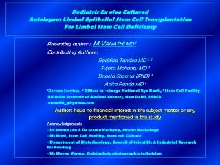
Pediatric Ex vivo Cultured Autologous Limbal Epithelial Stem Cell Transplantation For Limbal Stem Cell Deficiency Presenting author : M.VANATHI MD 1 Contributing Authors : Radhika Tandon MD 1, 2 Sujata Mohanty MD 3 Shweta Sharma (PhD) 3 Anita Panda MD 1 1Cornea Services, 2 Officer In -charge National Eye Bank, 3 Stem Cell Facility All India Institute of Medical Sciences, New Delhi, INDIA vanathi_g@yahoo.com Authors have no financial interest in the subject matter or any product mentioned in this study Acknowledgements: Dr Seema Sen & Dr Seema Kashyap, Ocular Pathology Ms Himi, Stem Cell Facility, Stem cell Culture Department of Biotechnology, Council of Scientific & Industrial Research for Funding Ms Meena Verma, Ophthalmic photographic technician
Pediatric Ex vivo Cultured Autologous Limbal Epithelial Stem Cell Transplantation For Limbal Stem Cell Deficiency • LSCD poor corneal epithelialisation persistent epithelial defects corneal vascularisation & scarring conjunctivalisation of the cornea Introduction Limbal Stem Cell Deficiency (LSCD) affects corneal epithelialisation resulting in persistent epithelial defects, which heal with corneal vascularisation, scarring, & conjunctivalisation 1. decreased vision ocular discomfort Symblepharon / ankyloblepharon restricted ocular motility unstable ocular surface / Dry eye syndrome Post chemical injury total LSCD Pellegrini et al 2 showed that • the corneal progenitor cells located in the limbus can be ex vivo expanded to generate cohesive sheets of corneal epithelium • Autologous cultured corneal epithelium can effectively restore the corneal surface • Dua HS, Azuara-Blanco A. Limbal stem cells of the corneal epithelium. Surv Ophthalmol. 2000 Mar-Apr; 44(5):415-25 • Pellegrini G, Traverso CE, Franzi AT, Zingirian M, Cancedda R, De Luca M. Long-term restoration of damaged corneal surfaces • with autologous cultivated corneal epithelium. Lancet. 1997 Apr 5; 349(9057):990-3.
Pediatric Ex vivo Cultured Autologous Limbal Epithelial Stem Cell Transplantation For Limbal Stem Cell Deficiency Purpose: To report the clinical outcome in ex vivo cultured autologous limbal stem cell transplantation for LSCD in children Methods: Retrospective analysis of ex vivo cultured autologous limbal epithelial stem cell (LESC) transplant done in children Clinical Evaluation & HPE examination Parameters noted: Demographic profile LSCD details (epithelial haze and irregularity, conjunctivalisation of cornea, & unstable ocular surface) Ocular surface evaluation (appearance of palisades of Vogt, presence of conjunctivalisation of tornea, symblepharon, tear break up time, Schirmer’s test ) histopathological examinations (morphology of epithelial cells and cells on impression cytology) Institutional Ethical Committee Approval was sought and obtained for the study Impressions Cytology shpwing (i) goblet cells (ii) normal corneal epithelium Diagnosis of LSCD
Pediatric Ex vivo Cultured Autologous Limbal Epithelial Stem Cell Transplantation For Limbal Stem Cell Deficiency Methods: Limbal Tissue Harvesting Limbal tissue specimens were obtained after a proper informed consent and approval. All Procedures were adhered to the guidelines of the Declaration of Helsinki in Biomedical Research Involving Human subjects. The autologous limbal tissue harvest was obtained (MV) from the patients in accordance to standard described techniques 3. Standardization of Human Limbal Explant Culture over Amniotic Membrane In a preliminary study the LESC culture standardization was done. • Denuded human amniotic membrane (dHAM ) was used for LESC expansion • Outgrowth of cells was observed from the explants within 1-2 days of culture, acheiving cell confluence in the 2nd week • Initial outgrowing cells were loosely adhered to HAM , & were spherical & translucent. Later adhered tightly to the membrane forming a dense honey comb like structure. At the end of 2 weeks cells formed a multilayer in culture. The expansion diameter was approximately 0.5 - 2.5cm. Ex vivo expanded LESC • Tsai RJ, Li LM, Chen JK. Reconstruction of damaged corneas by transplantation of autologous limbal epithelial cells. • N Engl J Med. 2000 Jul 13;343(2):86-93.
Pediatric Ex vivo Cultured Autologous Limbal Epithelial Stem Cell Transplantation For Limbal Stem Cell Deficiency Methods Surgical Procedure The exvivo cultured LESC was transplanted (MV) under GA Following a 360 0 peritomy, the fibrovascular pannus was excised by lamellar keratectomy The extent of the entire cornea was then covered by ex-vivo expanded LESC Fixation was done with 8-0 vicryl sutures or Human Fibrin glue (HFG) BCL was placed insitu for three weeks. Ex vivo cultured autologous LESC tranplant fixated with HFG
Pediatric Ex vivo Cultured Autologous Limbal Epithelial Stem Cell Transplantation For Limbal Stem Cell Deficiency Results Six eyes (6.2 + 2.6 years, range: 3 - 10 yrs, FU 9.8 + 7.5 months, range: 1.5 – 18 m) with LSCD underwent autologous ex vivo expanded LESC transplant. Postoperative phase showed (i) increased epithelial transparency (ii) in conjunctivalisation (iii) in superficial corneal vascularisation (iv) severity of symblepharon (v) mproved tear stability Corneal clarity improved in the initial postoperative period coinciding with time of best visual acuity Epithelial haze worsened later resulting in fall in visual acuity in late postoperative months. Improvement in ocular surface stability was seen in all cases CASE No 3 Postoperative (6 months later) Preoperative
Pediatric Ex vivo Cultured Autologous Limbal Epithelial Stem Cell Transplantation For Limbal Stem Cell Deficiency Results
Pediatric Ex vivo Cultured Autologous Limbal Epithelial Stem Cell Transplantation For Limbal Stem Cell Deficiency CASE No 6 Preoperative Note the LSCD with the lack luster appearance of the ocular surface Postoperative (6 months) Note the decrease in conjunctivalisation & density of corneal scarring, and improvement in symblepharon & surface wettablity Early postoperative Ex vivo cultured autologous limbal Epithelial Stem cell transplantaion
Pediatric Ex vivo Cultured Autologous Limbal Epithelial Stem Cell Transplantation For Limbal Stem Cell Deficiency Ex vivo Cultured LSCT DALK done 8 months later DALK (6 months postop)
Pediatric Ex vivo Cultured Autologous Limbal Epithelial Stem Cell Transplantation For Limbal Stem Cell Deficiency Conclusion Late postoperative epithelial haze may be due to the fact that transplanted LESC are being replaced by a rejuvenated host epithelium system. The epithelium produced by the replenished host LESC system perhaps mimics near normal morphology explaining the subsequent epithelial haze in the late postoperative phase. Prolonged time in achieving corneal clarity and amblyopia prevent optimal visual rehabilitation.
Pediatric Ex vivo Cultured Autologous Limbal Epithelial Stem Cell Transplantation For Limbal Stem Cell Deficiency • Exvivo Cultured Autologous LESC Transplantation helps in achieving • Improved ocular surface wetablity and stability. Conjunctivalisation reappeared in all cases though of lesser severity. The grade of severity of symblepharon improved • Postoperative appearance of palisades of Vogt supports the hypothesis 4 that the exvivo cultured LSCT aides in reviving the niche environment for revival of the damaged stems at the limbus • Epithelial haze was minimal in the early postoperative months (upto 3 – 6 months). Fall in visual acuity is attributed to the mild haze and astigmatism that develops due to the scarred ocular surfaces. Late postoperative epithelial haze may also be due to the fact that there is no longer any persistence of transplanted donor LESC on host ocular surface & that they are perhaps being replaced by a rejuvenated host epithelium system. • Shortt AJ, Secker GA, Notara MD, Limb GA, Khaw PT, Tuft SJ, Daniels JT. Transplantation of ex vivo cultured limbal epithelial stem cells: a review of techniques and clinical results. Surv Ophthalmol. 2007 Sep-Oct;52(5):483-502.
Pediatric Ex vivo Cultured Autologous Limbal Epithelial Stem Cell Transplantation For Limbal Stem Cell Deficiency Discussion Thorough scientific understanding of cultured LSCT is still evolving Only 2 – 9% of the entire limbal epithelial population in vivo comprises of LESC 5, 6. The exact percentage of stem cells present in exvivo cultured LESC sheets and behavior of post-transplant exvivo expanded LESC over a period of time are still largely unknown Reintegration of the cultured LESC onto the diseased ocular surface and their ability to replenish corneal epithelium remain the key in the success of exvivo LSCT Clinical evidence points to persistence of donor LESC on host ocular surface for about seven to nine months postoperatively and thereafter being replaced by host epithelium Our observations also support conclusions of Short et al 4 that exvivo cultured LESC transplantation does not provide a population of long term functional LESC but perhaps functions as a niche environment facilitating the revival of damaged host stem cell in the long term. • Budak MT, Alpdogan OS, Zhou M, Lavker RM, Akinci MA, Wolosin JM. Ocular surface epithelia contain ABCG2-dependent side population cells exhibiting features associated with stem cells. J Cell Sci. 2005 Apr 15;118(Pt 8):1715-24. • Watanabe K, Nishida K, Yamato M, Umemoto T, Sumide T, Yamamoto K, Maeda N, Watanabe H, Okano T, Tano Y. Human limbal epithelium contains side population cells expressing the ATP-binding cassette transporter ABCG2. FEBS Lett. 2004 May 7;565(1-3):6-10.