Download
1 / 42
460 likes | 1.08k Views
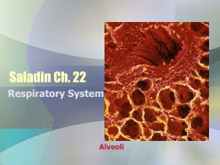
Respiratory Monitoring*. Jana A Stockwell, MD 2005. * Not vent waveforms or ABG analysis. Physical Exam. Monitor: Latin for “to warn” Observation : respiratory rate; pattern; color; nasal flaring; retractions; accessory muscle use Auscultation : wheeze; stridor; air entry; crackles; rales.

E N D
Respiratory Monitoring* Jana A Stockwell, MD 2005 * Not vent waveforms or ABG analysis
Physical Exam Monitor: Latin for “to warn” • Observation: respiratory rate; pattern; color; nasal flaring; retractions; accessory muscle use • Auscultation: wheeze; stridor; air entry; crackles; rales
Impedance Pneumography • 3 leads- • 1 over the heart • 2 on opposite sides of the lower chest • Small current is passed through 1 pair of electrodes • Impedance to current flow varies with the fluid content of the chest which, in turn, varies with the respiratory cycle • Converted into a displayed waveform
Pattern of breathing • Pause • Occurs in babies <3 mo, resolves by 6 months • Last <3 seconds • Occurs in groups of ≥3, separated by <20 sec • Apnea • NIH Conference consensus statement • Cessation of breathing for longer than 20 seconds or any respiratory pause associated with bradycardia, pallor or cyanosis
Pulse Oximetry • Non-invasively measures %HgbO2 • Beer-Lambert law: concentration of an unknown solute in a solvent can be determined by light absorption • Wavelengths of 660nm (red) and 940nm (infrared) • Absorption characteristics of the 2 hemoglobins are different at these 2 wavelengths
Pulse Oximetry • Correlates well, if true sat 70-100% ±2% of true sat 68% of time ±4% of true sat 96% of time • May not correlate with ABG sat • Several studies demonstrate that a fall in SpO2 often precedes any change in other VS
Pulse Oximetry - Mechanics • Light source is applied to an area of the body that is narrow enough to allow light to traverse a pulsating capillary bed and sensed by a photo detector • Each heartbeat results in a influx of oxygen saturated blood which results in increased absorption of light • Microprocessor calculates the amounts of HgbO2 and reduced Hgb to give the saturation
Functional vs Fractional • Pulse ox yields functional saturation • Ratio of HgbO2 to the sum of all functional hemoglobins (not CO-Hgb) • Sites filled/sites available for O2 to stick • Fractional saturation measured by co-oximetry by blood gas analysis • Ratio of HgbO2 to the sum of all hemoglobins
Absorption characteristics falsely account for a low sat in the patient with Hgb-Met Hgb-CO & Hgb-O2 have similar absorbance at 666nm so Hgb-CO will be falsely interpreted as Hgb-O2 (high sat)
Pulse Oximetry- Confounders • Misses other Hgb species (Hgb-CO, Hgb-Met) • Low perfusion states, severe edema or peripheral vascular disease make it difficult for the sensor to distinguish the true signal from background • Increased venous pulsations causes overestimation of deoxyHgb & decreased sats • Adversely affected by external light sources & motion artifact
Pulse Oximetry - Anemia Hgb 8, Sat 100% Decreased O2 content Until Hgb<5, then sampling errors Hgb 15, Sat 100% Normal O2 content
Transcutaneous • Developed in late 1970’s for use in neonates • Electrode is placed on a well-perfused, non-bony surface, skin is warmed to 41-44oC to facilitate perfusion and allow diffusion of gases • Estimates partial pressure of O2 & CO2 • Several studies demonstrated better oxygen correlation with pulse ox
CXR • Several studies in adults and pediatrics show significance of CXR to evaluate ETT or CVL location • One peds study showed that CXR was more sensitive than PEx for detecting significant problems • Consider routine use with infants or patients being proned
Capnography • Infrared spectroscopy • Compares the amount of infrared light absorbed to amount in chamber with no CO2 • Factors affecting : • Temp • Pressure • Presence of other gases • Contamination of sample chamber • Calibration
Capnography – Mainstream sampling • Advantages: • No aspiration of liquid • No lag time • No mixing gases in sample tube • Disadvantages: • Bulky airway adaptor • Must be intubated • Adds dead space • Moisture can contaminate chamber
Capnography – Sidestream sampling • Advantages: • Easier to calibrate • No added weight to airway • Less dead space • Less likely to become contaminated • Disadvantages: • Lag time for transit of sample • If TV small or flow rate high, inhaled gas may be aspirated with exhaled gas
Capnography • Best if… • Low flow sample rates • Fast response times • Improved moisture handling and purge systems • Calibration and correction for environmental factors
CO2 Physiology • CO2 transported in blood • 5-10% carried in solution reflected by PaCO2 • 20-30% bound to Hgb & other proteins • 60-70% carried as bicarbonate via carbonic anhydrase
CO2 Physiologya-ADCO2 • Normally 2-3mmHg • Widened if • Incomplete alveolar emptying • Poor sampling • High VQ abnormalities (normal 0.8), seen with PE, hypovolemia, arrest, lateral decubitus • Decreased with shunt • a-ADCO2 small • Causes: • Atelectasis, mucus plug, right mainstem ETT
CapnogramsNormal • Zero baseline • Rapid, sharp uprise • Alveolar plateau • Well-defined end-tidal point • Rapid, sharp downstroke A—B Deadspace B—C Dead space and alveolar gas C—D Mostly alveolar gas D End-tidal point D—E Inhalation of CO2 free gas
CapnographySudden loss of waveform • Esophageal intubation • Ventilator disconnect • Ventilator malfunction • Obstructed / kinked ETT
Sudden hypotension Massive blood loss Cardiac arrest Hypothermia PE CPB CapnographyDecrease in waveform
CapnographyGradual increase in waveform • Increased body temp • Hypoventilation • Partial airway obstruction • Exogenous CO2 source (w/laparoscopy/CO2 inflation)
CapnographySudden drop – not to zero • Leak in system • Partial disconnect of system • Partial airway obstruction • ETT in hypopharynx
Asthma PE Pneumonia Hypovolemia Hyperventilation CapnographySustained low EtCO2 Low ETCO2, but good plateau 40 30
CapnographyCleft in alveolar plateau • Partial recovery from neuromuscular blockade 40
CapnographyTransient rise in ETCO2 • Injection of bicarbonate • Release of limb tourniquet 40
CapnographySudden rise in baseline • Contamination of the optical bench – need to recalibrate 40
Question 1 1. State two ways oxygen is carried in the blood. a. Dissolved in plasma and bound with hemoglobin. b. Dissolved in plasma and bound with carboxyhemoglobin. c. Bound with hemoglobin and carbon monoxide. d. Dissolved in hemoglobin and bound with plasma.
Question 2 Which of the following statements about total oxygen content is true? a. The majority of oxygen carried in the blood is dissolved in the plasma. b. The majority of oxygen carried in the blood is bound with hemoglobin. c. Only 1% to 2 % of oxygen carried in the blood is bound with hemoglobin. d. Total oxygen content is determined by hemoglobin ability to release oxygen to the tissues.
Question 3 3. Which of the following statements about hypoxemia is false? a. Obstructive sleep apnea may cause carbon dioxide retention, but not hypoxemia. b. Certain postoperative patients are at greater risk for hypoxemia. c. Confusion may be a symptom of hypoxemia. d. Even the obstetric patient may be at risk for hypoxemia.
Question 4 Pulse oximetry incorporates two technologies that require: a. Red and yellow light. b. Pulsatile blood flow and light transmittance. c. Hemoglobin and methemoglobin. d. Veins and arteries.
Question 5 Which of the following defines “SpO2”? a. Partial pressure of oxygen provided by an arterial blood gas. b. Oxygen saturation provided by an arterial blood gas. c. Oxygen saturation provided by a pulse oximeter. d. Partial pressure of oxygen provided by a pulse oximeter.
Question 6 If your patient’s oxygen saturation has fallen from 98% to below 90%, after receiving 4 liters O2 via nasal cannula, the following physiologic changes may be occurring: a. Oxygen content is rapidly decreasing. b. PaO2 level is rapidly increasing. c. Oxygen content is slowly decreasing. d. PaO2 level is slowly increasing.
Question 7 Pulse oximetry can be used to: a. Obtain invasive information about oxygenation. b. Provide acid-base profiles. c. Noninvasively monitor saturation values during ventilator weaning. d. Fully replace arterial blood gas testing.
Question 8 Which of the following clinical conditions may contribute to inaccurate oxygen saturation readings as measured by a pulse oximeter? a. Venous pulsations. b. Mild anemia. c. Sensor placed on a middle finger. d. Monitoring a patient during weaning from oxygen.
Question 9 To troubleshoot motion artifact on a finger or toe sensor: a. Ensure the light source is directly across from the photodetector. b. Position the sensor below the level of the heart. c. Cover the sensor with an opaque material. d. Apply additional tape to the sensor to secure it in place.
Question 10 What is the PaO2 at 50% SpO2?: a. 88 b. 68 c. 48 d. 28