Download

1 / 19
690 likes | 4.94k Views
INVASIVE PRESSURE MONITORING. AIM Information concerning cardio respiratory performance and the effects of therapy in critically ill patients. 1. Arterial Blood Pressure Monitoring 2 . Central Venous Pressure Monitoring 3. Pulmonary Artery Pressure Monitoring

E N D
INVASIVE PRESSURE MONITORING AIM Information concerning cardio respiratory performance and the effects of therapy in critically ill patients
1. Arterial Blood Pressure Monitoring • 2 . Central Venous Pressure Monitoring • 3. Pulmonary Artery Pressure Monitoring • 4. Intra Cranial PressureMonitoring • 5. Intra Abdominal Pressure Monitoring
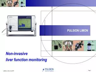
THINGS NEEDED • 1. PRESSURE TRANSDUCER • 2. DOME • 3. PRESSURE TUBINGS(arterial extension, 3 way stopcock, syringe) • 4. DISPLAYING UNIT • 5. TRANSDUCER STAND • 6. PRESSURE BAG WITH NORMAL SALINE • 7. CATHETER SET
Pressure transducer • Air mineture rugged and disposable device • These device can convert the movement of the sensing diaphargm into an electrical signal
Pressure tubings • The catheter and stopcock are normally attached to the flush device and transducer by non elastic pressure tubing • Continous flush device-use to fill pressure monitoring system and prevent from clotting into the catheter by continously flushing fluid through the system at the rate of 1 to 3 ml/hr
DISPLAY • Pressure waveforms are visualised on a calibrated oscilloscope • Digital displays provide a simple method for presenting quantitative data from the pressure waveform
CALIBRATION • The accuracy of blood pressure measurement requires an accurate reference point that is the patient mid axillary line • Zeroing process is done by closing the patient side and opening the other end of the three way to the atmosphere • Now press zero
ARTERIAL PRESSURE • INDICATIONS • Routine measurement of systemic blood pressure in icu • Multiple blood gas analysis PREFERABLE SITES • Radial,femoral,dorsalis pedis • CANNULATION • Collateral perfusion can be evaluated by modified allens test
CANNULE • 20G INSYTE (radial) • Single lumen 18G seldinger technique (femoral) • COMPLICATIONS • Infection • Thrombosis • Digital ischemia • Vessel damage • Bleeding, Nechrosis, Haemotoma
Indications 1.fliid administration 2.TPN hypertonic solutions 3.vasoactive infusions 4.monitoring for right atrial pressure(CVP) • PREPARABLE SITES 1.Internal jugular 2.subclavian 3.brachial 4.femoral
COMPLICATIONS 1.At insertion arterial puncture,pneumothorax,haemothorax 2.passage of wire/catheter arrhythmias,perforation of SVC,RA 3.Thrombosis 4.Catheter embolism
INDICATION • Haemodynamic measurement(cardiac output, stroke volume, systemic vascular resistance) • Measurement of right heart pressure(RAP,PAP) • Acute pulmonary hypertension • Pulmonary embolism • Cardiac tamponade • Estimation or preload/left heart filling(PAOP) • Derivation of oxygen variables(VO2,DO2)
HAEMODYNAMIC MEASUREMENTS 1.CARDIAC OUTPUT a.injectate 10ml 5% dextrose @ room temp b.inject througout the resp cycle c.>3 measurements and ignore values>10% from average 2.DERIVED VARIABLE a.CO/CI and SVR b.PVR(I), SV(I),LVSWI,RVSWI