Download
1 / 92
1.29k likes | 2.23k Views
Diagnosis and Management of Cerebral Venous Thrombosis

E N D
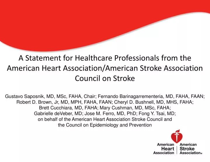
Diagnosis and Management of Cerebral Venous Thrombosis A Statement for Healthcare Professionals from the American Heart Association/American Stroke Association Council on StrokeGustavo Saposnik, MD, MSc, FAHA, Chair; Fernando Barinagarrementeria, MD, FAHA, FAAN; Robert D. Brown, Jr, MD, MPH, FAHA, FAAN; Cheryl D. Bushnell, MD, MHS, FAHA; Brett Cucchiara, MD, FAHA; Mary Cushman, MD, MSc, FAHA; Gabrielle deVeber, MD; Jose M. Ferro, MD, PhD; Fong Y. Tsai, MD; on behalf of the American Heart Association Stroke Council and the Council on Epidemiology and Prevention
AHA/ASA This slide set was adapted from the Diagnosis and Management of Cerebral Venous Thrombosis paper This statement reflects a consensus of expert opinion following thorough literature review that consisted of a look at clinical trials and other evidence related to the management of cerebral venous thrombosis. © 2011 American Heart Association, Inc. All rights reserved. Unauthorized use prohibited.
Applying classification of recommendations and levels of evidence. http://stroke.ahajournals.org/cgi/content/full/STROKEAHA.108.189696 © 2011 American Heart Association, Inc. All rights reserved. Unauthorized use prohibited.
Outline • Introduction • Epidemiology and Risk Factors for CVT • Clinical Diagnosis of CVT • Imaging in the Diagnosis of CVT • Management and Treatment • CVT in Special Populations • Clinical Outcomes: Prognosis • Summary © 2011 American Heart Association, Inc. All rights reserved. Unauthorized use prohibited.
Introduction • Thrombosis of the dural sinus and/or cerebral veins (CVT) is an uncommon form of stroke • CVT represents approximately 0.5-1% of all strokes • Multiple factors have been associated with CVT, but only some of them are reversible • This statement reviews the literature on CVT and provides recommendations for its diagnosis and management © 2011 American Heart Association, Inc. All rights reserved. Unauthorized use prohibited.
Epidemiology and Risk Factors • CVT is an uncommon and frequently unrecognized type of stroke that affects about 5 people per million annually and accounts for 0·5-1% of all strokes • CVT is more commonly seen in young individuals • According to the largest cohort study [International Study on Cerebral Venous and Dural Sinuses Thrombosis (ISCVT)], 487/624 (78%) occurred in patients younger than 50 © 2011 American Heart Association, Inc. All rights reserved. Unauthorized use prohibited.
Epidemiology and Risk Factors • Clinical features are diverse, and for this reason, cases should be sought among diverse clinical index conditions • No population studies have reported the incidence of CVT • In the Registro Nacional Mexicano de Enfermedad Vascular Cerebral (RENAMEVASC), a multihospital prospective Mexican Stroke Registry, 3% of all stroke cases were CVT © 2011 American Heart Association, Inc. All rights reserved. Unauthorized use prohibited.
Cortical veins 17% Superior sagital sinus Posterior frontal vein Trolar vein 62% Deep venous system 11% Anterior frontal vein Straight sinus 18% Transverse (lateral) sinus 41-45% Sigmoid sinus Internal Jugular Vein 12% © 2011 American Heart Association, Inc. All rights reserved. Unauthorized use prohibited.
Epidemiology and Risk Factors • A clinic-based registry in Iran reported an annual CVT incidence of 12.3 per million • In a series of intracerebral hemorrhage (ICH) in young people, CVT explained 5% of all cases • Predisposing causes of CVT are multiple • Risk factors are usually divided into acquired (eg. surgery, trauma, pregnancy, puerperium, antiphospholipid syndrome, cancer, exogenous hormones) and genetic (inherited thrombophilia) © 2011 American Heart Association, Inc. All rights reserved. Unauthorized use prohibited.
Clinical Diagnosis of CVT • The diagnosis of CVT is typically based on clinical suspicion and imaging confirmation • Clinical findings in CVT usually fall into two major categories, depending on the mechanism of neurologic dysfunction: • Those related to increased intracranial pressure (ICP) due to impaired venous drainage • Those related to focal brain injury from venous ischemia/infarction or hemorrhage © 2011 American Heart Association, Inc. All rights reserved. Unauthorized use prohibited.
Clinical Diagnosis of CVT • Many patients have clinical findings due to both mechanisms • Headache, generally indicative of an increase in ICP, is the most common symptom in CVT and was present in nearly 90% of patients in the ISCVT • Similar headache frequency has been reported in other populations studied © 2011 American Heart Association, Inc. All rights reserved. Unauthorized use prohibited.
Clinical Diagnosis of CVT • The headache of CVT is typically diffuse and often progresses in severity over days to weeks • A minority of patients may present with thunderclap headache, and migrainous type headache has been described • Isolated headache without focal neurologic findings or papilledema occurs in up to 25% of patients with CVT and presents a significant diagnostic challenge © 2011 American Heart Association, Inc. All rights reserved. Unauthorized use prohibited.
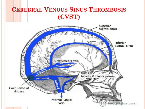
Clinical Diagnosis of CVT • Clinical manifestations of CVT may also depend on the location of the thrombosis • The superior sagittal sinus is most commonly involved and may lead to headache, increased ICP, and papilledema • For lateral sinus thromboses, symptoms of an underlying condition—middle ear infection—may be noted • Approximately 16% of CVT patients have thrombosis of the deep cerebral venous system (internal cerebral vein, Vein of Galen and straight sinus), which can lead to thalamic or basal ganglial infarction © 2011 American Heart Association, Inc. All rights reserved. Unauthorized use prohibited.
Clinical Diagnosis of CVT • Several clinical features distinguish CVT from other mechanisms of cerebrovascular disease • Focal or generalized seizures are quite frequent, occurring in about 40% of patients • An important clinical correlate to the anatomy of cerebral venous drainage is that bilateral brain involvement is not infrequent • Bilateral motor signs may also be present • CVT often presents with slowly progressive symptoms • In the ISCVT, onset to diagnosis was >48 hours to 30 days in 56% of patients © 2011 American Heart Association, Inc. All rights reserved. Unauthorized use prohibited.
Clinical Diagnosis of CVT:Recommendations Routine Blood Work • Class I Recommendations • In patients with suspected CVT, routine blood studies consisting of a complete blood count, chemistry panel, prothrombin time and activated partial thromboplastin time should be performed • Screening for potential prothrombotic conditions that may predispose CVT (i.e.: use of contraceptives, underlying inflammatory disease, infectious process, etc) is recommended in the initial clinical assessment (specific recommendations for testing for thrombophilia are found in the long-term management section of the main document) © 2011 American Heart Association, Inc. All rights reserved. Unauthorized use prohibited.
Clinical Diagnosis of CVT: Recommendations D-dimer Testing • Class II Recommendation • A normal D-dimer level using sensitive immunoassay or rapid Enzyme-Linked ImmunoSorbent Assay(ELISA) may be considered to help identify patients with low probability of CVT • If there is a strong clinical suspicion of CVT, a normal D-dimer level should not preclude further evaluation © 2011 American Heart Association, Inc. All rights reserved. Unauthorized use prohibited.
Clinical Diagnosis of CVT: Recommendations Intracranial Hemorrhage • Class I Recommendation • In patients with lobar intracerebral hemorrhage of otherwise unclear etiology or with cerebral infarction crossing typical arterial boundaries, imaging of the cerebral venous system should be performed © 2011 American Heart Association, Inc. All rights reserved. Unauthorized use prohibited.
Clinical Diagnosis of CVT: Recommendations Isolated Headache/Idiopathic Intracranial Hypertension • Class I Recommendation • In patients with the clinical features of idiopathic intracranial hypertension, imaging of the cerebral venous system is recommended to exclude CVT • Class II Recommendation • In patients with headache associated with atypical features, imaging of the cerebral venous system is reasonable to exclude CVT © 2011 American Heart Association, Inc. All rights reserved. Unauthorized use prohibited.
Imaging in the Diagnosis of CVT • Diagnostic imaging of cerebral venous thrombosis may be divided into two categories: non-invasive modalities and invasive modalities • The goal is to determine vascular and parenchymal changes associated with this medical condition • In some cases, the diagnosis is made only with cerebral digital subtraction angiography © 2011 American Heart Association, Inc. All rights reserved. Unauthorized use prohibited.
Imaging in the Diagnosis of CVT • Non-Invasive Diagnostic Modalities: Computed Tomography (CT), Magnetic Resonance Imaging (MRI) and Ultrasound • Invasive Diagnostic Angiographic Procedures: Cerebral Angiography and Direct Cerebral Venography © 2011 American Heart Association, Inc. All rights reserved. Unauthorized use prohibited.
Imaging in the Diagnosis of CVT • CT is widely used as the initial imaging test in patients presenting with new onset neurological symptoms • CT without contrast is often normal but may demonstrate findings suggesting CVT • The primary sign of acute CVT on a non-contrast CT is hyperdensity of a cortical vein and/or dural sinus • Only about one-third of CVT demonstrates direct signs of hyperdense dural sinus © 2011 American Heart Association, Inc. All rights reserved. Unauthorized use prohibited.
Non-contrast CT head scan showed hyperdensity of right transverse sinus as acute thrombosis © 2011 American Heart Association, Inc. All rights reserved. Unauthorized use prohibited.
Imaging in the Diagnosis of CVT • An ischemic infarction, sometimes with a hemorrhagic component, may be seen on CT • An ischemic lesion that crosses usual arterial boundaries (particularly with a hemorrhagic component) or in close proximity to a venous sinus, is suggestive of CVT • Subarachnoid hemorrhage and ICH are infrequent • Thrombosis of the posterior portion of the superior sagittal sinus may appear as a dense triangle, the dense or filled delta sign © 2011 American Heart Association, Inc. All rights reserved. Unauthorized use prohibited.
Imaging in the Diagnosis of CVT • Contrast-enhanced CT may show enhancement of the dural lining of the sinus with a filling defect within the vein or sinus • Contrast enhanced CT may show the classic “empty delta” sign - a central hypointensity due to very slow or absent flow within the sinus is surrounded by contrast enhancement in the surrounding triangular shape in the posterior aspect of the superior sagittal sinus © 2011 American Heart Association, Inc. All rights reserved. Unauthorized use prohibited.
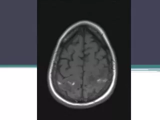
Imaging in the Diagnosis of CVT • In general, MRI is more sensitive for the detection of CVT than CT • The MR signal intensity of venous thrombus varies according to the time of imaging from the onset of thrombus formation • In the first week, venous thrombus is frequently isointense to brain tissue on T1 weighted image and hypointense on T2 weighted image due to increased deoxyhemoglobin • By the second week, thrombus contains methemoglobin, resulting in hyperintensity on T1 and T2 images © 2011 American Heart Association, Inc. All rights reserved. Unauthorized use prohibited.
Imaging in the Diagnosis of CVT • The principal early signs of CVT on non-contrast enhanced MRI are the combination of absence of a flow void with alteration of signal intensity in the dural sinus • MRI of the brain is suggestive of CVT by the absence of a fluid void signal in the sinus, T2 hypointensity suggestive of a thrombus, or a central isodense lesion in a venous sinus with surrounding enhancement • The secondary signs of MRI may show similar patterns to CT, including cerebral swelling, edema and/or hemorrhage • Brain parenchymal lesions of CVT are better visualized and depicted on MR than CT © 2011 American Heart Association, Inc. All rights reserved. Unauthorized use prohibited.
Flair MRI showed hyperintensity signal at left sigmoid sinus (arrow) © 2011 American Heart Association, Inc. All rights reserved. Unauthorized use prohibited.
T 2 weighted MRI showed high intensity bland venous infarct in frontal lobe © 2011 American Heart Association, Inc. All rights reserved. Unauthorized use prohibited.
Imaging in the Diagnosis of CVT • CT venography can provide a rapid and reliable modality for detecting CVT • CT venography is at least equivalent to MR venography in the diagnosis of CVT • Drawbacks to CT venography include radiation exposure, potential for iodine contrast allergy and use of contrast in the setting of poor renal function • In some settings, MR venography is preferable to CT venography due to these concerns © 2011 American Heart Association, Inc. All rights reserved. Unauthorized use prohibited.
Imaging in the Diagnosis of CVT • The most commonly used MR venographic techniques are time of flight (TOF) MR venography and contrast-enhanced MR venography • The 2D TOF (2 dimensional TOF) technique is the most commonly used method because 2D TOF has excellent sensitivity to slow flow compared with 3D TOF • Contrast enhanced MR venography offers improved visualization of cerebral venous structures © 2011 American Heart Association, Inc. All rights reserved. Unauthorized use prohibited.
MR venogram confirmed thrombosis (black arrows) of right transverse and sigmoid sinuses and jugular vein © 2011 American Heart Association, Inc. All rights reserved. Unauthorized use prohibited.
Imaging in the Diagnosis of CVT • Invasive cerebral angiographic procedures are less commonly needed to establish the diagnosis of CVT given the availability of MRV and CTV • These techniques are reserved for situations in which the MRV or CTV results are inconclusive or if an endovascular procedure is being considered © 2011 American Heart Association, Inc. All rights reserved. Unauthorized use prohibited.
Imaging in the Diagnosis of CVT • Cerebral Angiography • Findings include the failure of sinus appearance due to occlusion, venous congestion with dilated cortical, scalp or facial veins, enlargement of typically diminutive veins from collateral drainage, and reversal of venous flow • Acute dural sinus and cortical vein thrombosis typically cause a delay in cerebral venous circulation, and cerebral angiography will demonstrate delayed and slow visualization of cerebral venous structures © 2011 American Heart Association, Inc. All rights reserved. Unauthorized use prohibited.
Imaging in the Diagnosis of CVT • Direct Cerebral Venography • Direct cerebral venography is usually performed during endovascular therapeutic procedures • Intraluminal thrombus is seen either as a filling defect within the lumen in the setting of non-occlusive thrombosis or as complete nonfilling in occlusive thrombosis • Venous pressure measurements may be performed during direct cerebral venography to identify venous hypertension (normal venous sinus pressure is less than 10 mm of water) © 2011 American Heart Association, Inc. All rights reserved. Unauthorized use prohibited.
Venous phase of cerebral angiogram showed extensive thrombosed superior sagittal sinus and many frontal cortical veins © 2011 American Heart Association, Inc. All rights reserved. Unauthorized use prohibited.
Imaging in the Diagnosis of CVT: Recommendations • Class I Recommendations • Although a plain CT or MRI is useful in the initial evaluation of patients with suspected CVT, a negative plain CT or MRI does not rule out CVT. A venographic study (either CT or MR venogram) should be performed in suspected CVT if the plain CT or MRI is negative, or, to define the extent of CVT if the plain CT or MRI suggests CVT © 2011 American Heart Association, Inc. All rights reserved. Unauthorized use prohibited.
Imaging in the Diagnosis of CVT: Recommendations • Class I Recommendations • An early follow up CTV or MRV is recommended in CVT patients with persistent or evolving symptoms despite medical treatment or with symptoms suggestive of propagation of thrombus • In patients with previous CVT who present with recurrent symptoms suggestive of CVT, repeat CT or MR venogram is recommended © 2011 American Heart Association, Inc. All rights reserved. Unauthorized use prohibited.
Imaging in the Diagnosis of CVT: Recommendations • Class II Recommendations • Gradient Echo T2 susceptibility weighted images combined with MR venography can be useful to improve the accuracy of CVT diagnosis • Catheter cerebral angiography can be useful in patients with inconclusive CTV or MRV in whom a clinical suspicion for CVT remains high • A follow up CTV or MRV at 3-6 months following diagnosis is reasonable to assess for recanalization of the occluded cortical vein/sinuses in stable patients © 2011 American Heart Association, Inc. All rights reserved. Unauthorized use prohibited.
Management and Treatment • Organized care is one of the most effective interventions to reduce mortality and morbidity following acute stroke • CVT is an uncommon but potentially serious and life-threatening cause of stroke • Based on findings for stroke unit care in general, management of CVT in a stroke unit is reasonable for the initial management of CVT in order to optimize care and minimize complications © 2011 American Heart Association, Inc. All rights reserved. Unauthorized use prohibited.
Management and Treatment Initial Anticoagulation • There are several rationales for anticoagulation therapy in CVT: to prevent thrombus growth, to facilitate recanalization, and to prevent DVT or pulmonary embolism (PE) • Controversy has ensued because cerebral infarction with hemorrhagic transformation or intracerebral hemorrhage is commonly present at the time of diagnosis of CVT, and it may also complicate treatment © 2011 American Heart Association, Inc. All rights reserved. Unauthorized use prohibited.
Management and Treatment • Two available randomized controlled trials have compared anticoagulant therapy to placebo or open control in patients with confirmed CVT • One trial of 20 patients assessed intravenous unfractionated heparin (UFH) using dose adjustment to achieve an aPTT twice the pre-treatment value compared to placebo • The primary outcome was a CVT severity scale at 3 months, the secondary outcome was ICH • The trial was stopped early after 20 of a planned 60 patients were enrolled because there was a benefit of treatment © 2011 American Heart Association, Inc. All rights reserved. Unauthorized use prohibited.
Management and Treatment • The other trial of 59 patients compared subcutaneous nadroparin to placebo for 3 weeks followed by 3 months of oral anticoagulation (without placebo control) in those randomized to nadroparin • The study was blind during the first 3 weeks and open label thereafter • Primary outcomes were scores for activities of daily living, the Oxford Stroke handicap scale, and death • Secondary endpoints were symptomatic ICH and other major bleeding • At 3 months, 13% in the nadroparin group had a poor outcome compared to 21% with placebo (treatment difference in favor of nadroparin -7%; 95% CI -26 to 12%) © 2011 American Heart Association, Inc. All rights reserved. Unauthorized use prohibited.
Management and Treatment • Meta-analysis of these two trials revealed a non-statistically significant relative risk reduction of death or dependency with anticoagulation; RR 0.46 (95% CI 0.16 to 1.31), with a risk difference in favor of anticoagulation of -13% (95% CI -30 to 3%) • The relative risk of death was 0.33 (95% CI 0.08 to 1.21), risk difference -13% (95% CI -27-1%) © 2011 American Heart Association, Inc. All rights reserved. Unauthorized use prohibited.
Management and Treatment • A third trial randomized 57 women with puerperal CVT confirmed only by CT imaging and excluding those with hemorrhage on CT • Treatment was with subcutaneous heparin 5000 IU every 6 hours, dose adjusted to an aPTT 1.5 times baseline for at least 30 days after delivery • Outcome assessment was not blinded • Three patients in the control group either died or had residual paresis compared to none in the heparin group © 2011 American Heart Association, Inc. All rights reserved. Unauthorized use prohibited.
Management and Treatment • In the special situation of CVT with cerebral hemorrhage on presentation, even in the absence of anticoagulation, hemorrhage is associated with adverse outcomes • In one trial of nadroparin, all 6 deaths in the trial overall occurred in the group of 29 patients with hemorrhage on their pre-treatment CT scan • None of the deaths were attributed to new or enlarged hemorrhage • Cerebral hemorrhage was strongly associated with mortality, but not with cerebral bleeding on treatment © 2011 American Heart Association, Inc. All rights reserved. Unauthorized use prohibited.
Management and Treatment • A number of observational studies, both prospective and retrospective, are available, primarily from single centers • In a retrospective study of 102 patients with CVT, 43 had an ICH • Among 27 (63%) who were treated with dose-adjusted, intravenous heparin after the ICH, 4 died (15%), and 14 (52%) patients completely recovered • Mortality was higher (69%) with lower improvement in functional outcomes (3 patients completely recovered) © 2011 American Heart Association, Inc. All rights reserved. Unauthorized use prohibited.
Management and Treatment • The largest study by far was the ISCVT, which included 624 patients at 89 centers in 21 countries • Nearly all patients were treated with anticoagulation initially and mortality was 8.3% over 16 months • 79% had complete recovery (modified Rankin scale 0-1), 10.4% had mild to moderate disability (mRS 2-3) and 2.2% remained severely disabled (mRS 4-5) © 2011 American Heart Association, Inc. All rights reserved. Unauthorized use prohibited.
Management and Treatment • Few studies had sufficient numbers of patients not treated with anticoagulation to adequately address the role of anticoagulation in relation to outcome • Data from observational studies suggest a range of risks for ICH after anticoagulation for CVT from zero to 5.4% • In conclusion, limited data from randomized controlled clinical trials in combination with observational data on outcomes and bleeding complications of anticoagulation support a role for anticoagulation in treatment of CVT, regardless of the presence of pre-treatment ICH © 2011 American Heart Association, Inc. All rights reserved. Unauthorized use prohibited.
Management and Treatment Fibrinolytic Therapy • Although patients with CVT may recover with anticoagulation therapy, 9-13% have poor outcomes despite anticoagulation • Anticoagulation alone may not dissolve a large and extensive thrombus and the clinical condition may worsen even during heparin treatment • Incomplete recanalization or persistent thrombosis may explain this phenomenon © 2011 American Heart Association, Inc. All rights reserved. Unauthorized use prohibited.
Management and Treatment • Combining four studies including 114 CVT patients, partial or complete recanalization at 3-6 months was observed in 94 (82.5%) • Recanalization rates may be higher for patients receiving thrombolytic therapy • In general, thrombolytic therapy is used if clinical deterioration continues despite anticoagulation or if a patient has elevated intracranial pressure that evolves despite other management approaches © 2011 American Heart Association, Inc. All rights reserved. Unauthorized use prohibited.