Download
1 / 6
60 likes | 91 Views
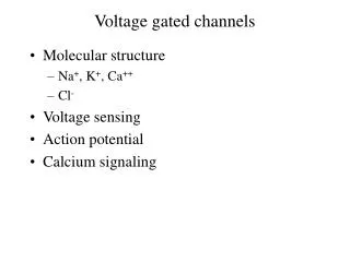
This text provides insights into the activation states of voltage-gated ion channels including Ca2+, Cl-, K+, and Na+ channels, as well as Ligand-Gated Ion Channels like nAcChR, Serotonin, and GABAAR. It delves into the structural compositions and activation processes of various ion channels, highlighting the conformational changes and pore formations upon activation. Additionally, it covers the structures of Calcium, Potassium, Sodium channels, and HCN Channel, explaining the functional units and subunit arrangements in each type of ion channel.

E N D
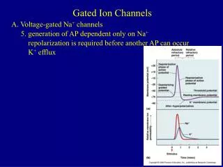
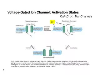
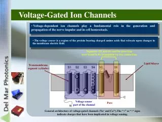
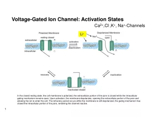
Voltage-Gated Ion Channel: Activation States Ca2+,Cl-,K+, Na+-Channels Li+ In the closed resting state, the cell membrane is polarized, the extracellular portion of the pore is closed while the intracellular gating mechanism remains open. Upon activation, the membrane depolarizes, opening the extracellular portion of the pore and allowing the ion to enter the cell. The refractory period occurs while the membrane is still depolarized, the gating mechanism has closed the intracellular portion of the pore, rendering the channel inactive.
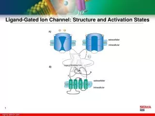
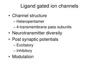
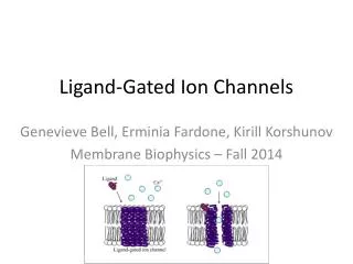
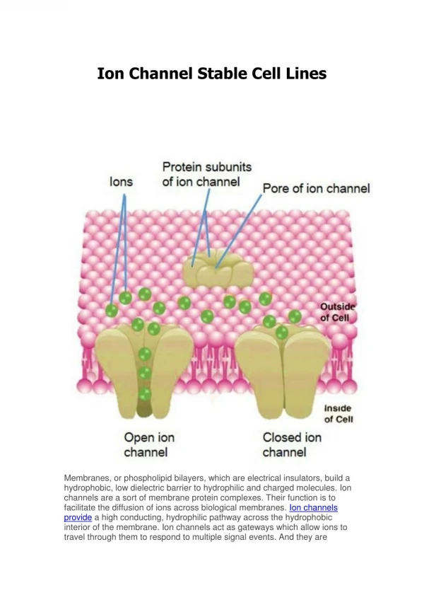
Ligand-Gated Ion Channel: Structure and Activation States nAcChR, Serotonin, GABAAR, incl. Li+ • The intrinsic ion channel in this superfamily of receptors undergoes a conformational change following the binding of an extracellular ligand to its binding site, which allows for the opening of the channel pore. • The pore is formed by a pentameric arrangement of subunits, which are in turn composed of a large extracellular domain (which contains the ligand binding site) and four transmembrane domains.
Calcium Channel Structure Li+ • The a1 subunit is comprised of four homologous domains, each of which contains six transmembrane helices. The b subunit is intracellular and associates with the a1 subunit. The g subunit is a glyoprotein that possesses four transmembrane segments. The a2 subunit is extracellular, highly glycosylated and associates with the membrane spanning d subunit via disulfide bonds. • The four domains of the a1 subunit cluster in the membrane to form the pore region.
Schematic Representation of the HCN Channel -Hyperpolarization-Activated Cyclic Nucleotide-Gated- Li+ Cation-Channel (Na+ ,K+)
2TM Potassium Channel Structure 6TM Potassium Channel Structure Li+ Li+ Four of these subunits cluster to form the active channel. Each subunit is composed of two membrane-spanning helices connected by a P loop. • A) The a subunit is formed from 6 transmembrane segments and is associated with a regulatory b subunit. • B) Four a subunits form the pore.
Sodium Channel Structure Li+ A) The a subunit consists of four domains of six transmembrane segments, with the 5th and 6th segments comprising the pore formation unit. The b subunits consist of a single membrane spanning segment with an immunoglobulin-like fold in an extracellular domain. The a subunit contains the binding sites for a variety of toxins and drugs, as indicated. B) The four domains of the a subunit form the pore within the membrane.