Download
1 / 26
270 likes | 468 Views
Voltage gated channels. Molecular structure Na + , K + , Ca ++ Cl - Voltage sensing Action potential Calcium signaling. Core voltage-gating functional unit. 6 transmembrane segments One charged Pore facing Ion selectivity & V-dependence Tetrameric organization

E N D
Voltage gated channels • Molecular structure • Na+, K+, Ca++ • Cl- • Voltage sensing • Action potential • Calcium signaling
Core voltage-gating functional unit • 6 transmembrane segments • One charged • Pore facing • Ion selectivity & V-dependence • Tetrameric organization • 4x separate, 6 pass proteins • 1 protein with 4, 6 pass domains Transmembrane domain Potassium channel has 4 separate subunits PDB: 2r9r
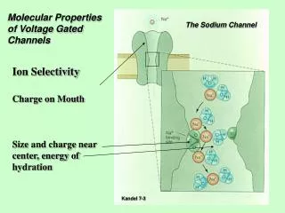
Voltage gated sodium channel Sodium channel has 4 functional domains • Ion selectivity and voltage sensitivity from S4 helices • Long cytoplasmic loops btw domains • Intracellular domains subject to modification • Conductivity • Open probability
Domain organization Organization • Common prokaryotic ancestor • S5-S6 • 4 subunit/domain • Pore forming motif K+ structure Canonical subunit S2 S1 S5-6 S3 S4
Voltage sensing • Transmembrane potential stabilizes S4 • S4 moves S5/S6 • Pore open/close 6 1 4 2 5 3
Chloride Channel • Double barreled, 2 subunit channel • Each subunit has 3 charged helices with anti-parallel arrangement forming V-sensor PDB: 1kpl
Applied V Current Time Recorded I Whole cell recording State Model • Clamp voltage • Record current • Aggregate channel activity & density • G=1/R=I/V C O V Derived Conductance Derived I-V Rectification: Current diverges from straight-line conductance
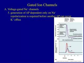
Channel Inactivation State Model • Feedback mechanism • Membrane depolarization • Reduces driving force • Secondary conformational change C O V I Channel becomes refractory with depolarization Channel opens with depolarization Depolarization Voltage steps Preconditioned Depolarized
Characteristics of voltage gated channel • Conductance • Ion selectivity • Threshold • Open time • Inactivation time
Anatomy of Action potential • Voltage gated channels selectively drive intracellular potential between different ionic equilibrium potentials • K+ -90mV • Na+ +60mV Threshold for V-gated Na+ channels Neural AP Cardiac AP
Ionic currents in AP Step voltage to increasing depolarization Net current Na+ current Sub-threshold Depolarizing current Inactivates K+ current Large depolarization opens new K+ channels “Delayed rectifier”
Ionic currents in AP Current declines over time, even though potential remains constant
Ionic contributions to AP • Kleak (Kir) set resting potential • Inactivate at threshold • NaV • Open at threshold • Rapid, large g • KV • Open at threshold • Delayed rectifier (slow) • Large g
Anatomy of Cardiac AP • Leaky membranes (7) give slow depolarization to • Threshold opens CaV (3) & NaV (1) • KV (4) and KCa repolarize • Prolonged AP vs neuron • Ca current • Much delayed K+
Na+ NaV causes local depolarization • Membrane capacitance of 10-6 F/cm2 • 10-6 (p r2) • Na influx: n (1.6 10-19 C) • Threshold ~-40mV • V=Q/C • 20-30 channels/micron2 • ~ 400 ions/channel to depolarize neighbors Na+ r -40 mV -90 mV
Cm Cm Equivalent Circuit • Borrowed from cable theory • Break cell into parallel compartments • Propagation depends on resistance/capacitance Extracellular Rm Rm Cm Cm Ri Intracellular
Neural cable theory • Neuron size vs conduction velocity • Large diameter, low internal resistance • Myelinated/Unmyelinated • Insulates membrane • Increases RM • Decreases CM • Increase V • Node of Ranvier
NaV Modulation • 10 genes • Alternative splicing • Phosphorylation • Protein binding • Alters • Threshold • Conductivity • Kinetics • Selectivity PKA PKC PKA PKC Cn RPTP Cn RPTP Kinases decrease conduction Phosphatases increase conduction • -28 identified binding partners • Cytoskeletal • Adhesion • Signaling
Calcium channel • Most common effector of AP • Same basic structure as other VG channels • Major classes • N-type “Neuronal” • L-Type “Long” • T-Type “Tiny”
Neurons • Ionotropic = channels • Metabotropic = receptors • Neurotransmitter release depends on [Ca2+]I • Multiple inputs Nerve terminals & presynaptic vessicles
N-type calcium channels • Neurotransmitter release (presynaptic) • Calcium dynamics same time scale as firing (10 ms) • Highly localized changes (50-5000 nm) • Post-synaptic, Ca-dependent remodeling
Striated Muscle • Cardiac • Skeletal • “Twitch” force • 50-200 ms • All-or-none • Tension depends on [Ca2+]I • Spontaneous • Neural
L-type calcium channels • Excitation contraction coupling • Long open time (100 ms) • Modulation • Calcium dependent inhibition • Oxidation • Phosphorylation
T-Type calcium channels • Tiny conductance (6 vs 25 pS) • Low threshold (-50 vs -30 mV) • Regulatory role • Cell differentiation • Modulation of phenotype • Neuronal bursting
Smooth muscle • Tonic • Vascular • Respiratory • Phasic • GI • Bladder • Tension depends on [Ca2+]I • Hormonal • Mechanical • Neural Smooth muscle cells in vasculature, gut, sphincter
Smooth Muscle Calcium • Ligand gated Ca channels • Voltage gated Ca channels • Second messengers