Glomerulonephritis
Glomerulonephritis . Dr. Abdelaty Shawky Dr. Gehan mohamed. Learning objectives. Classification of glomerulonephritis. Pathological features of minimal change glomerular disease. Pathological features of post-streptococcal glomerulonephritis. Nephritic syndrome. Nephrotic syndrome.

Glomerulonephritis
E N D
Presentation Transcript
Glomerulonephritis Dr. Abdelaty Shawky Dr. Gehan mohamed
Learning objectives • Classification of glomerulonephritis. • Pathological features of minimal change glomerular disease. • Pathological features of post-streptococcal glomerulonephritis. • Nephritic syndrome. • Nephrotic syndrome.
Glomerular diseases constitute some of the major problems in nephrology; indeed, chronic glomerulonephritis is one of the most common causes of chronic renal failure in humans. • Glomeruli may be injured by a variety of factors and in the course of a number of systemic diseases. Systemic immunologic diseases such as systemic lupus erythematosus (SLE), hypertensionand polyarteritisnodosa, diabetes mellitus, often affect the glomerulus. • These are termed secondary glomerular diseases to differentiate them from primary renal disorders in which the kidney is the only or predominant organ involved.
* Classification of glomerular diseases: I. Primary G.N (the disease affects kidney only): • Minimal change glomerular disease (Lipoid nephrosis). • Acute diffuse proliferative G.N: • Post-streptococcal G.N. • Non-post-streptococcal GN. • Rapidaly progressive G.N. • MembraneousG.N. • MembranoproliferativeG.N. • Chronic G.N.
II. Secondary G.N (the disease affects kidney and other organs): • Systemic lupus erythematosus (SLE). • Polyarteritisnodosa (PAN). • Wegener granulomatosis. • Diabetes mellitus (diabeteic nephropathy). • Goodpasture syndrome. • Amyloidosis.
Most of the 1ry glomerular disease are due to immunologic mechanisms. • To study any glomerular disease, a renal biopsy is taken and examined by 3 types of microscopes: 1. Light microscope: to examine the structure of glomeruli, tubules and interstitium. 2. IF (immune flourescentmicroscope): to detect the type of deposited immunoglobulin in the glomeruli. 3. EM (electron microscope): to detect the site of immune complex, either sub-epithelial, sub-endothelial, mesangial or basement membrane..
*Etiology & pathogenesis: • Chemical change in the glomerular basement membrane causing protein loss.
* Grossly: • Mild bilateral kidney enlargement. * LM (Light microscope): • No abnormalities. * IF (Immunoflurescence): • No immune deposits. * EM (Electron microscope): • Fusion of the foot processes of the epithelial cells (podocytes).
* CP (Clinical picture): • Affect children and young adults. • Cause nephrotic syndrome. * Fate: • The disease has excellent prognosis and most patients respond to corticosteroids with complete resolution of proteinuria.
*Etiology & pathogenesis: • Immune complex reaction; (nephrotegenic strains of group A beta haemolytic streptococci + Ig G), the complex is deposited in the glomeruli with subsequent complement activation acute inflammation.
* Grossly: • Mild bilateral kidney enlargement with petechial hemorrhages.
* LM (Light microscope): a. Glomeruli: • Proliferation of endothelial and mesangial cells. • increase number of neutrophils inside Glomerular capillaries . • Bowman’s space shows: neutrophils, RBCs, some albumin. b. Tubules: • The lining cells are swollen. • The lumens show casts (RBCs casts, neutrophil casts & hyaline casts). c. Interstitium: • Acute inflammatory reaction…...
* IF (Immunoflurescence): • Deposition of IgG and C3.
* EM (Electron microscope): • Subepithelial immune complex deposit (humps).
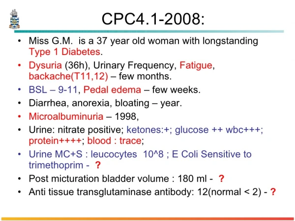
* CP (Clinical picture): • In the classic case, a young child abruptly develops malaise, fever, nausea, oliguria, and hematuria (smoky or cocoa-colored urine) 1 to 2 weeks after recovery from a sore throat. • The patients exhibit red cell casts in the urine, mild proteinuria (usually less than 1 mg/day), peri-orbital edema, and mild to moderate hypertension.
Hematuria (coca cola colored urine) RBCs cast
In adults, the onset is more likely to be atypical, with the sudden appearance of hypertension or edema, frequently with elevation of serum creatinine. Important laboratory findings include elevations of anti-streptococcal antibody (ASO) titers and a decline in the serum concentration of C3 (consumed).
More than 95% of affected children eventually recover totally with conservative therapy aimed at maintaining sodium and water balance. • A small minority of children (perhaps less than 1%) do not improve, become severely oliguric, and develop a rapidly progressive glomerulonephritis. • Some of the remaining patients may undergo slow progression to chronic glomerulonephritis.
In adults, the prognosis is bad. Most of the patients pass to rapidly progressive glomerulonephritis or chronic renal failure.
Nephritic syndrome - A syndrome formed of: 1. Haematuria. 2. Oliguria. 3. Peri-orbital oedema. 4. Hypertension. - The most common cause of nephritic syndrome in children is post-streptococcal GN.
Nephrotic syndrome - A syndrome formed of: 1. Hypoproteinaemia. 2. Proteinuria . 3. Oedema. 4. Hypercholesterolaemia. • The most common cause of nephrotic syndrome in children is minimal change glomerular disease. • The most common cause of nephrotic syndrome in adults is membranous GN.
Thanks References: Robbins and Cotran’s: Pathologic Basis of Disease. Seventh edition.