Download
1 / 12
150 likes | 664 Views
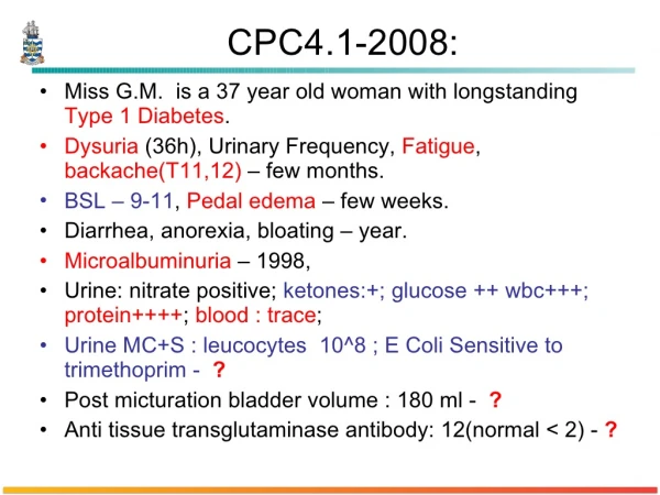
Acute Glomerulonephritis. Investigations. Urinalyis MSU Biopsy. Urinalysis. Urine is dark . Specific gravity is greater than 1020 osm . Proteinuria is observed . Haematuria RBCs and red cell casts are present on microscopy ( P erform FBE to check for anaemia ) Leukocytes

E N D
Acute Glomerulonephritis Investigations
Urinalyis • MSU • Biopsy
Urinalysis • Urine is dark. • Specific gravity is greater than 1020 osm. • Proteinuriais observed. • Haematuria • RBCsand red cell casts are present on microscopy • (Perform FBE to check for anaemia) • Leukocytes • Creatinine level increased
Asymptomatic urinary abnormalities • Isolated proteinuria without haemturia • May be early sign of glomerular lesion- i.e. membranous GN, IgA nephropathy, diabetic nephropathy or amyloidosis • Haematuria with/without sub-nephrotic range proteinuria • SLE, Henoch-Schonleinpurpura, post-infectious GN
Blood • FBE • ESR, CRP for inflammation • U + E’s • LFT’s • Serum albumin (low in nephrotic syndrome) • Glucose- to exclude diabetes • Serum complement (low in SLE) • Auto-antibodies: serum immunoglobulins, ANCA (Wegener’s granulomatosus, anti-ss-DNA (SLE)
Imaging • Chest radiography • needed in patients with a cough, with or without haemoptysis(ie, Wegener granulomatosis, Goodpasture syndrome, pulmonary congestion). • Abdominal CT is needed if visceral abscesses are suspected; also look for chest abscesses. • Echo • For pts with new cardiac murmur or a positive blood culture • rules out endocarditis or a pericardial effusion. • Renal US: • to evaluate kidney size as well as to determine the extent of fibrosis. • A kidney size of less than 9 cm is suggestive of extensive scarring and a low likelihood of reversibility • CT scan of the head for patient with malignant hypertension or altered mental status.
Biopsy • Candidates for biopsy: • individual or family history of renal disease, atypical presentation: • massive proteinuria • nephrotic syndrome • rapid rise in creatinine level without resolution.
Post-strep GN • Biopsy shows diffuse, florid, acute inflamm at the glomerulus • No necrosis, but occaisonal crescents • Neutrophils and deposition of IgG and complement • Light microscopy of biopsy sample shows acute inflammation of glomerulus with neutrophils
IgA nephropathy (commonest form of GN) • Elevated IgA complex in mesangium • Biopsy shows mesangial cell proliferation and increased matrix
Wegener’s granulomatosus • C- ANCA that reacts with proteinase 3