GLOMERULONEPHRITIS
GLOMERULONEPHRITIS. Division of Nephrology, Internal Medicine. Symptoms and Signs. Injury to the glomeruli by various immunologic or non-immunologic mechanism P roteinuria H ematuria E dema H ypertension A zotemia. 사구체신염의 분류. 임상 증상에 따른 분류 ( 5 Syndromes)

GLOMERULONEPHRITIS
E N D
Presentation Transcript
GLOMERULONEPHRITIS Division of Nephrology, Internal Medicine
Symptoms and Signs Injury to the glomeruli by various immunologic or non-immunologic mechanism • Proteinuria • Hematuria • Edema • Hypertension • Azotemia
사구체신염의 분류 임상 증상에 따른 분류 ( 5 Syndromes) 1) 무증상성요이상(AUA) : IgA N 2) 신증후군(NS) : MCD, FSGS, MGN 3) 급성 사구체신염(AGN) : PSGN 4) 만성 사구체신염(CGN) : IgA N, MPGN 5) 급속진행성 사구체신염(RPGN) : crescentic GN
원인 질환에 따른 분류 1) 속발성사구체신염 HS nephritis, lupus nephritis, DM nephropathy Hepatitis B virus associated GN, PSGN 2) 원발성사구체신염 MCD, FSGS, MGN, MPGN, IgA N, Crescentic GN 병리학적(형태학적) 분류
NS Definition Children : Serum albumin < 2.5g/dl Proteinuria > 40mg/m2/hr Adult : Massive Proteinuria ( >3g/24h/1.73m2) Hypoalbuminemia Edema Hyperlipidemia
NS Anti-proteinuric Barrier • Charge selective barrier : negatively charged sialoglycoprotein • Size selective barrier : 44A(effective molecular radius)
NS Classification • Primary NS : MCD FSGS MGN MPGN • Secondary NS : Hepatitis B virus-associated GN Lupus nephritis, Diabetic nephropathy
NS Primary NS according to the age groups Others FSGS MGN MCD
NS Etiologic Diagnosis of Secondary NS (N=220) Diabetic nephropathy : m/c Others Renal amyloidosis 5.9% HBGN 46.8% Lupus Nephritis 39.5%
Normal Kidney (1) H&E stained section
Minimal Change Disease (MCD) • Synonyms : Nil disease, Lipoid nephrosis Foot process disease • Incidence : male>female 80% of NS in children 20-40 % of NS in adults • Cause : Drug (NSAIDs, rifampin, interferon a) Hodgkin’s disease, HIV infection • Pathology : normal in LM, IF epithelial foot process effacement in EM
MCD Treatment • Highly steroid responsive, excellent prognosis to glucocorticoid therapy 90% (children) and 50% (adult) : remission after 8weeks of high dose steroid therapy • sometimes relapse upon tapering (50-70%) • Alkylating agents are reserved for frequent relapsers, cyclosporinefor steroid resistant MCNS
MCD LM EM
FSGS • 특발성 신증후군의 10% 정도 차지, • 원인 : 특발성, heroin abuse, VUR, AIDS, • solitary kidney • 임상 양상 : 대부분 전형적인 신증후군(nonselective) • 고혈압, 혈뇨, 신기능 저하 • 병리 : 비증식성, 경화성 변화 • (초점성, 분절성) • 면역 형광 소견은 비특이적 • 치료 : 장기간의 steoid 치료, • steroid 에 대한 효과가 낮음 • 예후 : 50% 환자가 7-10년에 ESRD 로 진행 , • 신이식 후에도 재발 흔함
FSGS LM
MGN 1. Most common idiopathic NS in adults (40%) Rare in children Peak incidence between 30 and 50 2. Clinical manifestation Mostly nephrotic (80%), Nonselective proteinuria Microhematuria (50%) HT : less than 30% at initial manifestation but common later with renal progression
MGN Pathology LM : diffuse thickening of the GBM (PAS staining) spike pattern (Silver staining) IF : granular deposit of IgG, IgM, C3 EM : subepithelial electron dense deposit stage I, II, III, IV
MGN Etiology Idiopathic (majority) Systemic disease or drugs 1. Infection : hepatitis B, hepatitis C 2. Autoimmune disease : SLE, RA, MCTD 3. Malignancy : carcinoma 4. Drugs : gold, penicillamine, NSAID, captopril 5. Miscellaneous : sarcoidosis, DM,
MGN Clinical Sx ①Mean onset age: 30 ∼ 50 age (male: female = 2:1) ②Older age: correlate with malignancy 20% (>60 age) ③Massive proteinuria (80%), edema ④ RVT*: 50% - high incidence
MGN Treatment & Prognosis 40% : spontaneous remission 30-40% : repeated relapse and remission 10-20% : persistent NS and progressive azotemia (ESRD in 20 to 30 years) Risk factors of renal progression : male, older age at onset, heavy proteinuria, hypertension, stage IV lesion, azotemia at initial Bx Tx : no therapeutic effect with steroid alone cyclophosphamide, chlorambucil, cyclosporine
MGN LM
MGN IF EM IF with antibody to IgG. deposition of electron dense material and interposition of lighter GBM material-subepithelialdeposition
Type I: Subendothelial Deposits Type II: Dense Deposit Disease Membranous Proliferative Glomerulonephropathy
Type I: Subendothelial Deposits Type II: Dense Deposit Disease MPGN EM
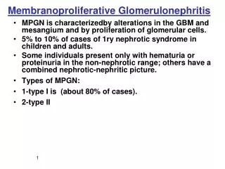
MPGN Clinical Manifestation Variable combination of nephritic or nephrotic features Common in ages between 5-30 Decline in GFR, active urine sediment, Proteinuria often in nephrotic range (50%) Type I MPGN : Immune complex disease C3 usually depressed C1q, C4, properdin, factor B : borderline or low Secondary MPGN : associated with infection, SLE, malignancy
MPGN Pathology Diffuse proliferation of mesangial cells Increased mesangial matrix Thickening and reduplication of GBM (Double contour, Tram-tract) Type I and type II MPGN Prognosis Poor, slow progression to ESRD Worse in type II MPGN
MPGN LM Normal kidney
MPGN EM IF
Acute Glomerulonephritis or Acute Nephritic Syndrome(AGN)
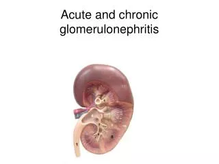
AGN Acute glomerular inflammation Sudden onset of acute renal failure and oliguria Obstruction of glomerular capillary lumen GFR falls Na and water retention ECF volume expansion, Edema, Hypertension U/A : RBC cast, dysmorphic RBC, leukocytes, subnephroticproteinuria Often gross hematuria Azotemia General pathologic feature : proliferative GN (capillary endothelial cell, mesangial cell)
PSGN AGN - PSGN Acute post-streptococcal GN Etiology : Pharyngeal or cutaneous infection with group A beta-hemolytic streptococcus, nephritogenic strain Epidemiology : common in children, male > female Latent period : 6-15 days (“post-pharyngitic”) Hematuria (gross or microscopic), Edema, mild HT, Oliguria, Nausea, mild fever, Flank pain ARF of variable degrees
PSGN Lab finding & Pathology U/A : hematuria, mild proteinuria GFR reduced Elevated ASO, anti-hyaluronidase, elevated anti-DNase B Culture: Streptococcus in throat or skin Complement level : C3, CH50 markedly reduced, normalized in 8 weeks, C4 mildly reduced LM : Diffuse endocapillary proliferation PMN cell and monocyte infiltration EM : hump (large subepithelial deposit: characteristic) IF : IgG, C3 deposit
PSGN Treatment & prognosis Benign course in children Acute symptom : relieved in 1-2 wks U/A abnormality esp. hematuria persists for 2 years Treatment : symptomatic Rest, salt water restriction, diuretics, protein restriction Antibiotics (PCN, EM) needed in limited cases - i.e. incomplete treatment, elevated CRP
PSGN LM IF
PSGN EM
Asymptomatic Urinary Abnormality (AUA) • Hematuria or subnephrotic proteinuria without HT, renal insufficiency, edema • U/A abnormality : persistent or recurrent
IgAN Most common GN worldwide (10-40%) Etiology : most cases are idiopathic Clinical spectrum with Henoch-Schonlein Purpura Secondary IgA N due to liver cirrhosis, Crohn’s disease Epidemiology : between 16 and 35 years, male>female Initial manifestation Recurrent gross hematuria, often 24 to 48h after pharyngitis, GI infection (“synpharyngitic”) Or, microscopic hematuria during routine examination HT, nephrotic proteinuria rare at initial presentation