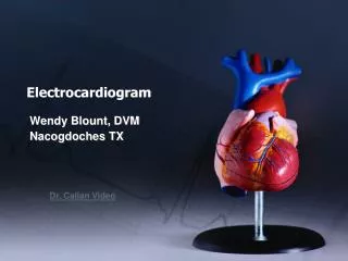
ELECTROCARDIOGRAM
ELECTROCARDIOGRAM. YINGMIN-CHEN Renji Hospital affiliated to Shanghai Second Medical University. WPW Diagram. ECG Intervals and Waves. ECG Components Diagram. RV vs LV PVC's. Diagram: AV Nodal Reentrant Tachycardia. Diagram: Type I vs. Type II 2nd Degree AV Block. QRS Axis = +90 degrees.


ELECTROCARDIOGRAM
E N D
Presentation Transcript
ELECTROCARDIOGRAM YINGMIN-CHEN Renji Hospital affiliated to Shanghai Second Medical University
Wandering Atrial Pacemaker Wandering atrial pacemaker is a benign rhythm change where the pacemaker site shifts from the sinus node into the atrial tissues. P-wave morphology varies with the pacemaker site.
PAC's with RBBB Aberrant Conduction PAC's are identified by the arrows. Note that the PP interval surrounding the PAC is less than 2x the basic sinus cycle indicating that the sinus node has been reset by the ectopic P wave. The pause after the PAC, therefore, is incomplete
Atrial Parasystole In atrial parasystole non-fixed coupled PACs, shown by arrows, occur at a common inter-ectopic interval or at multiples of this interval. Atrial fusions, not shown here, may also occur when the PAC occurs in close temporal proximity to the sinus impulse
In ventricular parasystole, non-fixed coupled PVC's occur at a common inter-ectopic interval. Fusion beats, indicated by arrows, are often seen. Fusions occur when the sinus impulse entering the ventricles find the ventricles already partially depolarized by the parasystolic focus
Ventricular Fusion Beats Fusion beats occur when two or more activation fronts contribute to the electrical event. These may occur in the atria or in the ventricles. In this example the ventricular fusions are the result of simultaneous activation of the ventricles from two foci, the sinus node and a ventricular ectopic focus
Nonconducted PACs and Junctional Escapes Although at first glance this looks like 2nd degree AV block, the P waves indicated by the arrows are premature and not sinus P waves. The pause is long enough to encourage a junctional escape focus to take over. Note the sinus P waves just before the escape beats. Had the escapes not occurred, the sinus impulses would have captured the ventricles
Nonconducted And Conducted PAC's The pause in this example is the result of a nonconducted PAC, as indicated by the first arrow. The second arrow points to a conducted PAC. The most common cause of an unexpected pause in rhythm is a nonconducted PAC
Identification of PVC's and PAC's PVC's usually stick out like sore thumbs; PAC's are often difficult to see because they are hidden in the preceding ST-T wave. The PVC in this example is mostly negative in lead V1 suggesting RV origin; i.e., most of activation is moving in posterior dirction towards the left ventricle
Nonconducted PAC's: An Unusual Bigeminy Occasionally nonconducted PAC's can create interesting rhythms. In this example every other sinus beat is followed by an early, nonconducted PAC. The resulting pause sets up a bigeminal rhythm. Note the distortion of the T waves caused by the nonconducted PAC's
An Interpolated PAC Although most PAC's reset the sinus node producing an "incomplete compensatory pause", this PAC, indicated by the black arrow, is interpolated, i.e., sandwiched between two sinus beats. Note that the subsequent sinus P wave conducts with prolonged PR interval due to the relative refractoriness of the AV junction left by the PAC. Auscultation of the heart during this single PAC event would reveal three rapid beats in a row, suggesting a brief tachycardia
These PAC's, indicated by arrows, enter the ventricles and find the right bundle refractory. They therefore conduct with RBBB aberrancy. In most normal hearts the right bundle recovery time is longer than the left bundle's; most aberrancy, therefore, has a RBBB morphology. In some diseased hearts the left bundle may have a longer refractory period resulting in LBBB aberration. Aberrant conduction may also involve the fasicles of the left bundle
Sore Thumbs Two funny looking premature beats are seen in this rhythm strip. Beat 'A' is preceded by a PAC which distorts the T wave, making this an aberrantly conducted PAC. Beat 'B' is a PVC. The notch on the downslope of the QRS complex clearly dentifies this as a PVC and not aberrancy