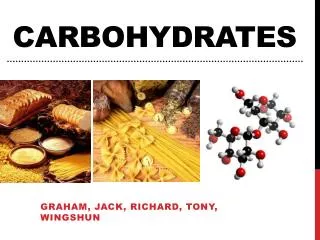
CARBOHYDRATES
CARBOHYDRATES. Medical Biochemistry Molecular Principles of Structural Organization of Cells. CARBOHYDRATES Are hydrated carbon molecules [C n H 2n O n or (CH 2 O) n ], They are virtually ubiquitous because they have such a wide range of structures and functions Structure:

CARBOHYDRATES
E N D
Presentation Transcript
CARBOHYDRATES Medical Biochemistry Molecular Principles of Structural Organization of Cells
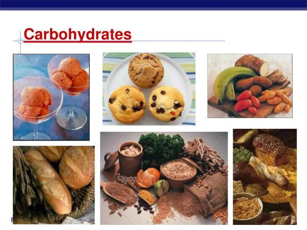
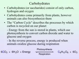
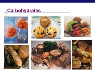
CARBOHYDRATES • Are hydrated carbon molecules [CnH2nOn or (CH2O)n], • They are virtually ubiquitous because they have such a wide range of structures and functions • Structure: • polyhydroxylated ketones, • polyhydroxylated aldehydes, or • compounds that can be hydrolyzed into these compounds. • A few of the functions of carbohydrates include the following. • provide the majority of energy in most organisms (simple carbohydrates are sugars; complex carbohydrates can be broken down into simple sugars). • provide the C atoms necessary for synthesis of lipids, proteins, nucleic acids • enter in the structure of complex compounds: • mucopoliglucides, • glycolipids, • coenzymes, • comprise large portions of the nucleotides that form DNA and RNA (ribose, deoxyribose) • serve as metabolic intermediates (glucose-6-P, fructose-1,6-bisP). • give structure to cell walls (in plants - cellulose) and cell membranes • play a role in lubrication, cellular intercommunication, immunity.
CARBOHYDRATE CLASSIFICATION AND NOMENCLATUREA.Classification • MONOSES (Monosaccharides) such as glucose and fructose, are simple sugars. They can be connected by glycosidic linkages to form more complex compounds, glycosides • COMPLEX GLUCIDES: • Homoglucides • Oligosaccharides, such as blood group antigens, are polymers composed of 2-10 monosaccharide units. For example: Disaccharides, such as maltose and sucrose, can he hydrolyzed to 2 monoses, trisaccharides to 3 monoses, tetrasaccharides to 4 monoses,… • Polysaccharides, such as starch and cellulose, are polymers composed of >10 monosaccharides. • Heteroglucides are formed of one carbohydrate and a noncarbohydrate component
CARBOHYDRATE CLASSIFICATION AND NOMENCLATUREB. Nomenclature • 1. Carbon numbering system. Monosaccharides are named according to a system that uses the number of carbons as the variable prefix followed by -ose as the suffix. • In the general formula CnH2nOn, nis the number of carbons. a. Triose = 3 carbons b. Tetrose = 4 carbons c. Pentose = 5 carbons d. Hexose = 6 carbons The carbons are numbered sequentially
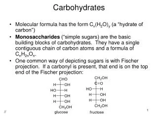
CARBOHYDRATE CLASSIFICATION AND NOMENCLATUREB. Nomenclature • 2. Reactive groups. The reactive group (aldehyde or ketone) on a carbohydrate determines whether it is an aldose or a ketose. • Aldoses are monosaccharides with an aldehyde (-CH=O) group as the reactive group (e.g. glucose). • Ketoses are monosaccharides with a ketone (>C=O) group as the reactive group (e.g. fructose). The aldehyde or ketone group is on the carbon with the lowest possible number • Monosaccharide and reactive-group nomenclature can be combined to designate compounds. For example, the sugar glucose is an aldohexose = a six-carbon monosaccharide (-hexose) containing an aldehyde group (aldo-).
THE CLASSIFICATION OF THE CARBOHYDRATES ALDOSES - C = O functional H carbonyl group KETOSES > C = O MONOSES TRIOSES (C = 3) glyceraldehyde, (MONOSACCHARIDES, number TETROSES (C = 4) SIMPLE SACCHARIDES of carbon PENTOSES (C = 5) ribose, deoxyribose SIMPLE GLUCIDES) atoms HEXOSES (C = 6) glucose,galactose,fructose HEPTOSES (C = 7) MONOSES DERIVATIVES CARBOHYDRATES (SUGARS, URONIC ACIDSAMINOGLUCIDESPHOSPHOESTERS SACCHARIDES, (glucuronic, (glucosamine (glucose-6-phosphate, GLUCIDES) galacturonic) galactosamine) fructose-1,6-diphosphate) DIGLUCIDES maltose, lactose, sucrose OLYGOGLUCIDES TRIGLUCIDES [2-6(10) monoses] TETRAGLUCIDES HOMOGLUCIDES etc. (HOMOGENEOUS POLYGLUCIDES Starch, CARBOHYDRATES) (10 monoses) Glycogen (only oses) Cellulose COMPLEX CARBOHYDRATES HETEROGENEOUS Mucopolyglucides, CARBOHYDRATES Glycolipids, (carbohydrate component + Glycoproteins, noncarbohydrate component) etc.
STRUCTURES – OPEN CHAIN FORMS • Monoses are • polyhydroxylated ketones • polyhydroxylated aldehydes • Isomers are compounds with the same chemical formula but with different structural formula • Function isomers: glucose aldehyde function and fructose keto function • Optical isomers (D and L) or enantiomers • Epimers are two isomers with conformations that are different only at one carbon atom.
All monosaccharides (simple sugars) contain at least one asymmetric carbon (a carbon bonded to four different atoms or groups of atoms). In glucose, carbons 2—5 (C2—C5) are asymmetric. Because of this carbon asymmetry, the sugars are optically active, and are namedenantiomers: • Configuration. The simplest carbohydrates are the trioses, such as glyceraldehyde, which has two optically active forms designatedL and D • Nomenclature. For the purposes of nomenclature, other sugars are considered to be derived from glyceraldehyde. Thus, a D-sugar is one that matches the configuration of D-glyceraldehyde around the asymmetric carbon that is the farthest from the aldehyde or ketone group. An L-sugar correspondingly matches L-glyceraldehyde. L-glyceraldehyde D-glyceraldehyde
Enantiomers are isomers that are mirror images. • As mirror images, enantiomers rotate the same plane of polarized light to exactly the same extent, but they do this in opposite directions, when they are in aqueous solution. L-glucose D-glucose • They have identical physical properties except for the direction of rotation of plane-polarized light. • If a plane of polarized light is rotated to the right (clockwise), the compound is dextrorotatory. • If a plane of polarized light is rotated to the left (counterclockwise), the compound is levorotatory.
Epimers are two isomers with conformations that are different only at one carbon atom. • Glucose and Mannose are epimers at C2 • Glucose and Galactose are epimers at C4 mannose glucose galactose
STRUCTURE – CYCLIC FORM • In aqueous solution monoses exist in chain form or a spontaneous reaction takes place between one of the hydroxyl groups and the carbonyl group leading to cyclic structures • five members = 4 carbon atoms and 1 oxygen atom (furanose) • six members = 5 carbon atoms and 1 oxygen atom (pyranose) Pentoses, such as ribose, form a five-membered ring (ribofuranose) Hexoses, such as glucose or galactose, form a five-membered ring (glucofuranose) or six-membered ring (glucopyranose)
Hemiacetals can occur in linear or cyclic forms. When an alcohol reacts with an aldehyde, linear, unstable compounds occur: intermolecular hemiacetals • Cyclic hemiacetals are formed by similar intramolecular reactions. In glucose, the hydroxyl group on C-5 can react intramolecularly with the carbonyl group on C-1 to form a stable cyclic hemiacetal.
Anomeric carbon is the new asymmetric carbon (C-1 in glucose) that is created by cyclization at the carbon bound to oxygen in hemiacetal formation, with essential role in reducing properties of glucides.a. If the hydroxyl on the anomeric carbon is below the plane of the ring, it is in the α position. b. If the hydroxyl on the anomeric carbon is above the plane of the ring, it is in the βposition. • Mutarotation is the process by which α and βsugars, in solution, slowly change into an equilibrated mixture of both. 1. α-D-Glucopyranose (62%); 2. β-D-Glucopyranose (38%); 3. α-D-Glucofuranose (trace); 4. β-D-Glucofuranose (trace); 5. Linear D-Glucose (0.01%).
Glucose α-D-glucopyranose β-D-glucopyranose α-D-glucofuranose β-D-glucofuranose
Galactose α-galactopyranose β-galactopyranose
Fructose α-fructofuranose β-fructofuranose
GLYCOSIDIC LINKAGES • A sugar can react with an alcohol to form an acetal known as a glycoside. • If the sugar residue is glucose, the derivative is a glucoside; • if the residue is fructose, the derivative is a fructoside. • a residue of galactose results in a galactoside derivative. • When the side chain (R) is another sugar, the glycoside is a disaccharide. e.g. maltose = α-D-glucopyranosyl-α-D-glucopyranoside sucrose = α-D-glucopyranosyl-β-D-fructofuranoside • If R is already a disaccharide, the glycoside is a trisaccharide and so forth.
TRIOSES glyceraldehyde dihydroxyacetone • Result as intermediary metabolites (in phosphoric esters form) in the reactions of carbohydrate degradation (glycolysis)
PENTOSES • Exogenous origin (food) • In the cell, have higher metabolic stability than hexoses • D-ribose (anomer β): • Does not exist free in the cell • Biological importance: as phosphate ester enters in the structure of nucleosides, nucleotides, RNA, coenzymes, metabolic intermediates in pentose-phosphate cycle • 2-Deoxy-D-ribose (anomer β) • In the structure of deoxyribonucleosides and nucleotides, structural monomers of deoxyribonucleic acid (DNA) β-D-ribose β-2-deoxy-D-ribose
HEXOSES • Aldohexoses • glucose = Glc = G (dextrose, blood sugar, grape sugar), • galactose = Gal (cerebrose), • mannose = Man • Ketohexose • fructose = Fru, F (levulose, fruit sugar)
- In the blood it exists in a constant interval of 65-110 mg/dl (glycemia); maintained mainly by the antagonistic action of 2 pancreatic hormones: • insulin - hypoglicemiant • glucagon – hyperglycemiant • The increased values of glycemia are present in diabetes mellitus and endocrine diseases GLUCOSE (Glc, G) • Ubiquitous in the animal and plant organisms • The main ose in the human organism • Location • In all the cells and fluids of the organism except the urine • Functions • energetic: through degradation (glycolysis) energy is generated as ATP • it enters in the structure of • diglucides: maltose, isomaltose, lactose, sucrose, celobiose • polyglucides: starch, glycogen, cellulose • by oxidation in the liver it is transformed in glucuronic acid with important role in detoxifying the organism.
GALACTOSE (Gal) • Location: it exists in reduced amount in blood, CSF, urine • Function: • With glucose forms lactose, the sugar in the milk • Enters in the structure of complex lipids in the brain (cerebrosides, sulfatides, gangliosides) • By oxydation in the liver forms the galacturonic acid that enters in the structure of mucopolyglucides (complex carbohydrates)
FRUCTOSE (Fru, F) • The sweetest of all sugars • Structure: ketohexose • pyranose in free form and • furanose in all natural derivatives • Location: • free in the secretion of seminal vesicles • combined with glucose forms the sucrose, the sugar in the fruits • as phosphoric ester is an intermediate in the metabolism of glucose (glycolysis and pentose-phosphate cycle),
CARBOHYDRATES DERIVATIVES 1. URONIC ACIDS • Are produced by the oxydation of the aldehyde carbon, the hydroxyl carbon or both • Glucuronic acid (GlcA, GlcUA) • pyranose form in natural products • component of proteoglycans • process of detoxification of normal biological compounds, waste products or toxins (phenols, alcohols, amines, amides, etc) • Galacturonic acid (GalA, GalUA) • component of glucosaminoglycans • components of pectins, plant gums, mucilages • in the bacterial polysaccharides
CARBOHYDRATES DERIVATIVES 2. AMINOSUGARS / AMINOGLUCIDES A hydroxyl group is replaced with amino or acetylamino group • D-glucosamine (GlcN, chitosamine) as • N-acetylglucosamine (GlcNAc) is the product of the hydrolysis of hyaluronic acid and chitin, the major component of the shells of insects and crustaceans; heparin, blood-group substances • N-acetyl-muramic acid is part of the bacterial membrane • D-galactosamine as • D-galactosamine sulfate found in polysaccharides of cartilage, chondroitin sulfate, • N-acetyl galactosamine • D-mannosamineas N-acetyl-neuraminic acid (AcNeu, NeuAc,sialic acid) is an essential component of the glycoproteins and glycolipids in the brain, erythrocyte stroma, bacterial cell membrane
CARBOHYDRATES DERIVATIVES 3. PHOSPHORIC ESTERS • Are formed from the reaction of phosphoric acid with a hydroxyl group of the sugar. Phosphorylation is the initial step of the metabolism of sugars. • They are metabolic intermediates • Examples: • glyceraldehyde-3-P, dihydroxyacetone-1-P, dihydroxyacetone-3-P
ribose-5-P ribose-3,5-bisP • glucose-1-P glucose-6-P • fructose-1-P fructose-1,6-bisP
BIOCHEMICAL IMPORTANCE OF MONOSES • Source of energy in the presence or absence of oxygen (aerobic or anaerobic glycolysis) • Plastic function as they are involved as derivatives in the buildup of diverse biological molecules (nucleosides, nucleotides, coenzymes, glycolipids, glycoproteins)
OLYGOSACCHARIDES / OLYGOGLUCIDES • Are complex glucides resulting from the condensation of 2-6(10) identical oses • Depending on the number of oses they can be: disaccharides (2 oses), trisaccharides (3 oses), tetrasaccharides (4 oses)… • Depending on the mechanism of water elimination they can have reducing properties or not: • Reducing disaccarides are formed when the molecule of H2O is eliminated between the hemiacetalic –OH of one ose and an alcoholic –OH of the second ose; the hemiacetalic or hemiketalic –OH of the second ose rests free. This type of bond is called monocarbonylic or glycosidic bond oriented α or β (e.g. maltose, isomaltose, lactose, cellobiose) • Nonreducing disaccharides are formed by the elimination of H2O between the two -OH hemiacetalic or hemicetalic, blocking both reducing groups in the bond (e.g. sucrose)
REDUCING DISACCHARIDES 1. MALTOSE • Structure: • It results from the condensation of 2 α-glucose • The bond in maltose is between C1 and C4 (α-1,4-glycosidic configuration) • It possesses an unattached anomeric carbon atom, thus it is a reducing sugar. • Role: • It exists in the structure of starch and glycogen from the food, resulting from their partial hydrolysis, catalyzed by the amylase from saliva and pancreatic juice • It is hydrolyzed in the intestine, under the action of maltase
2. ISOMALTOSE Structure: It results from the condensation of 2 α-glucose The bond is between C1-C6 (α-1,6-glycosidic) It possesses a free hemiacetalic –OH, thus it is a reducing sugar In the structure of amylopectin and glycogen 3. CELLOBIOSE Structure: It results from the condensation of 2 β-glucose The bond is between C1-C4 (β-1,4-glycosidic) The free hemiacetalic β-OH gives reducing properties It results from cellulose hydrolysis It is hydrolyzed in the digestive tract of herbivorous catalyzed by cellobiase produced by the microflora REDUCING DISACCHARIDES
REDUCING DISACCHARIDES 4. LACTOSE • milk sugar, slightly sweet • Structure: • formed of β-Galactose and α-Glucose • bond between C1-C4 (β-1,4) • α-OH hemiacetalic is free (reducing) • Synthesized by the mammary glands • Exists in milk as free diglucide (2-8%) • Its hydrolysis is catalyzed by lactase, in the intestine; the β-Gal is absorbed and transported to the liver where it is converted in α-Glu; if the enzyme is deficient the Gal is accumulated (galactosemia = genetic disease)
NONREDUCING DISACCHARIDES SUCROSE • In contrast to the linkages in most other simple carbohydrates, the oxygen bridge between α-Glucose and β-Fructose is between the hemiacetalic –OH at C1 of Glc and hemicetalic –OH at C2 of Fru (α,β-1,2-glycosidic linkage). • Consequently, there is no free hemiacetalic or hemicetalic -OH group in sucrose.Therefore, this disaccharide is not a reducing sugar. For example, it will not reduce an alkaline copper reagent such as Fehling’s solution. • Exists in the sugar beet and cane; it is very soluble • Its hydrolysis catalyzed by sucrase generates the 2 oses
POLYSACCHARIDES/POLYGLUCIDES/GLYCANS • Classification: • Homoglycans – products of polycondensation of one type of ose: • glucose → glycans : starch, glycogen, cellulose • galactose → galactosans • mannose → mannans, • arabinose →arabinans. • Heteroglycans – products of polycondensation of more types of structural units: • Mucopolyglucides components of proteoglycans • Bacterial polyglucides
HOMOGENEOUS POLYGLUCIDES • Result of the condensation of a great number of identical oses • The repeating unit is • maltose in starch and glycogen • cellobiose in cellulose • Role: reserve of energy • Structure: linear or branched • The hydrolysis catalyzed by hydrolases = glycosidases results in the component oses • Properties: • Hydrophilic - when placed in water they swell and then dissolve to form colloidal solutions, very viscous, capable of gelation
HOMOGENEOUS POLYGLUCIDES 1. STARCH • is the storage form of glucose in plants, resulting from photosynthesis • is formed of grains with characteristic microscopic appearance for each plant • has amorphous structure, is insoluble in water; in hot water forms a paste • has weak reducing properties • is identified in reaction with iodine (blue colour) • the enzyme catalyzed hydrolysis is progressive, generating intermediates with smaller molecular mass (dextrines) that have specific colours in reaction with iodine: → amylodextrines (blue-violet) → erythrodextrines (red) → flavodextrines (yellow) → acrodextrines (colorless) → maltose → glucose • the repeating unit is maltose • the grains are formed of amylose (20%) in the center and amylopectin (80%) as an envelope
Amylose • It is a linear unbranched polymer (M=105) formed of 100-400 α-glucose moieties (as maltose) linked with α-1,4-glycosidic bonds. • The chain has α-helix configuration (6 glucose each turn) • It has hemiacetal –OH only at the end of the chain (weak reducing properties) • It is soluble in hot water forming coloidal solution; in cold water forms a gel • It is identified in reaction with iodine (blue colour)
Amylopectin • It is a branched polymer (M=106-107) of α-glucose (10 000) linked with glycosidic bonds of 2 types: • α-1,4-glycosidic linkages (maltose type) and • α-1,6 (isomaltose type) branching points that occur at intervals of approximately • 16 α-D-glucose residues on the external chain and • 10 residues on the internal chain • The hydrolysis in the digestive tract implies the catalytic activity of: • α-amylase (salivary and pancreatic) acts on α-1,4 bonds in the middle of the chain → dextrines → maltose → glucose • 1,6-α-glycosidase acts on α-1,6 bonds → amylose • maltase acts on maltose → 2 α-glucose (absorbed in the intestine wall and transported to the liver)
HOMOGENEOUS POLYSACCHARIDES 2. GLYCOGEN • It is the major storage form of carbohydrate in animals (liver and muscle). • It is a highly branched form of amylopectin (M=106-107): • α-1,4-glycosidic linkages • α-1,6 branching points occur every 6-7 α-glucose residues in the external and 3 residues in the inner chains. • The hydrolysis of exogeneous glycogen is similar with the one of starch. • The endogeneous glycogen is transformed by: • phosphorolysis, catalysed by phosphorylase that act on α-1,4 bonds beginning with the nonreducing end of the chain → G-1-P: • In the liver, G-1-P is used to maintain the glycemia constant • In the muscle G-1-P → G-6-P used to provide the energy necessary for muscular contraction (glycolysis) • α-1,6-glycosylase that act on α-1,6 bonds.
HOMOGENEOUS POLYSACCHARIDES 3. CELLULOSE • It is a structural polysaccharide of plant cells (M= 106). • It is composed of linear (unbranched) chains of β-glucose units (cellobiose) joined by β-1,4-glycosidic linkages. • The chains can form fibers • The hydrolysis of β-1,4-glycosidic bonds is catalysed by cellulase or cellobiase that do not exist in the human digestive tract • Although cellulose forms a part of the human diet (in vegetables, fruit), only a very small amount is transformed under the action of the intestinal microflora • It is important for the maintenance of the intestinal movements, as a protective mean against the cancer of the colon.
HETEROGLUCIDES/GLYCOSAMINOGLYCANS (GAGs)/PROTEOGLYCANS • Structure: • glucide component (C,H,O, N and/or S) 85-90% of molecular mass and • non glucide component (protein) in small amount, • linked by covalent or electrovalent bonds with the proteins (proteoglycans), except the hyaluronic acid (only polyglucide) • they form viscous solutions, mucus • the name mucopolyglucides refers to heteropolyglucides of animal origin • Classification: depending on the nature of glucide component : • acidic = hexozamine + uronic acid • neutral = only hexozamine
Acidic GAGs • Structure: long unbranched polysaccharides containing repeating disaccharide units that contain hexosamine + uronic acid • The physiologically most important Acidic GAGs are • hyaluronic acid, • chondroitin sulfate, dermatan sulfate • heparin, heparan sulfate. • Location: found in the lubricating fluid of the joints and as components of cartilage, synovial fluid, vitreous humor, bone, and heart valves.
1. Hyaluronic acid • Structure: polyglucide macromolecule • Glucuronic acid + N-acetylglucozamine (β-1,3-bonds) = hyalobiuronic acid • The repeated units are linked β-1,4-bonds • Location: embryonic tissue, conjunctive tissue, cartilage, cornea, vitreous fluid, synovial fluid, umbilical cord • Role: tissue cement, lubricant, shock protective; marked capacity for binding water • Biosynthesis in the fibroblasts in 2 days • Depolymerized by hyaluronidase, • that acts on β-1,4-bonds • exists in the spermatozoa cap, venom, bacteria • In the tissues there is an anti-hyaluronidase (Physiologic Hyaluronidase Inhibitor = PHI)
2. Chondroitin sulfates • Structure: it is a polyglucide macromolecule atached to protein, composed of : • β-D-glucuronate + N-acetylgalactosamine-4-sulfate or 6-sulfate (linkedβ-1,3) = chondrosine • The units are linked β-1,4 • Location: cartilage, bones, tendons, skin, aorta, cornea • The great number of negative charges = cations changing resins, regulating the cartilage matrix structure and the storage of minerals in the bone matrix • They are attached to proteins and associated with hyaluronic acid forming supra-molecular complexes
Dermatansulfate • Chondroitinsulfate B (CSA-B) = dermatansulfate contains iduronic acid instead of GlcUA • Location: derm, tendons, heart valves, blood vessels. • When there is a deficiency of the lysosomal enzymes, they are unable to completely decompose the mucopolyglucides, thus the dermatansulfate is accumulated in the tissues, and excreted in the urine (Hurler disease - fatal)
3. Heparin • Structure: • A complex mixture of linear polysaccharides: • The diglucide units are varied (glucuronic or iduronic sulfated acid + glucozamine N-sulfated or N-acetylated) linked α-1,4. The degree of sulfation of the saccharide units is varied. • Location: in the blood, aorta, lungs • Synthesized in the mast cells lining the artery walls in the liver, skin, lungs • Role: • has anticoagulant properties and • coenzyme in lipoproteinlipase system from the walls of capillaries (role in the hydrolysis of triglycerides, VLDL)
NEUTRAL GAGsKeratansulfates • Formed of acetilated hexozamines, complexed with proteins • Location: cartilages associated with chondroitinsulfates, skin, conective tissue
FUNCTIONS OF SULFATED PROTEOGLYCANS • Binds water – in the tissues exposed to high pressures (joints cartilage, nucleus pulposus, skin) • Filter – salts and compounds with low molecular mass can diffuse (basement membranes) • Ionized at neutral pH – cation exchanger (Na+ is more concentrated in the matrix of cartilage) • Regulating calcification of cartilage, inhibiting the crystallization of calcium phosphate • Interact with fibrous proteins collagen or elastic, • Dermatansulfate, heparansulfate and heparine form insoluble complexes with LDL – involved in atherosclerosis patogenic mechanism • Heparin highly negatively charged, cannot coilup and cross-link; stable complexes with cations • Blood coagulation