Download
1 / 56
560 likes | 584 Views
Explore the nature of mutations, from DNA changes to protein alterations, and their impact on human health. Learn about germline vs. somatic mutations, associated diseases like sickle cell anemia and Ehlers-Danlos syndrome, and how mutations cause various illnesses. Discover the causes of mutations, including spontaneous and induced factors, and classification methods like point mutations.

E N D
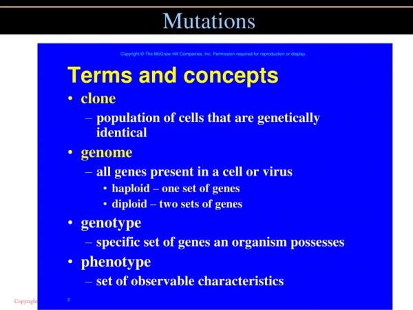
The Nature of Mutations A mutation is change in a DNA sequence that is present in < 1% of a population May occur at the DNA or chromosome level A polymorphism is a genetic change that is present in > 1% of a population The effect of mutations vary “Loss-of-function” mutations – Recessive “Gain-of-function” mutations – Dominant
The Nature of Mutations The term mutant refers to phenotype - Usually connotes an abnormal or unusual, or even uncommon variant that is nevertheless “normal” Mutations are important to evolution Our evolutionary relatedness to other species allows us to study many mutations in non-human species
The Nature of Mutations Germline mutations - Originate in meiosis - Affect all cells of an individual Somatic mutations - Originate in mitosis - Affect only cells that descend from changed cell
Mutations Alter Proteins Identifying how a mutation causes symptoms has clinical applications Examples of mutations that cause disease: - Beta globin gene - Collagen genes
Sickle Cell Anemia Results from a single DNA base change in the b-globin gene, which replaces glutamic acid (6th position) with valine Phenotype associated with homozygotes Altered surface of hemoglobin allows molecules to link in low oxygen conditions Creates sickle shape of RBC Sickling causes anemia, joint pain, and organ damage when RBC become lodged in small blood vessels
Collagen A major component of connective tissue - Bone, cartilage, skin, ligament, tendon, and tooth dentin More than 35 collagen genes encode more than 20 types of collagen molecules Mutations in these genes lead to a variety of medical problems
Collagen has a precise structure - Triple helix of two a1 and one a2 polypeptides - Longer precursor, procollagen is trimmed to form collagen Figure 12.3
Figure 12.4 Ehler-Danos Syndrome A mutation prevents procollagen chains from being cut Collagen molecules cannot assemble, and so skin becomes stretchy Figure 12.4
How Mutations Cause Disease Mutations in a gene may cause either different versions of the same disease or distinct illnesses Table 12.2 lists several examples of mutations and the diseases they produce Figure 12.4
Causes of Mutations Mutations may occur spontaneously or by exposure to a chemical or radiation An agent that causes a mutation is called a mutagen Figure 12.4
Spontaneous Mutation De novo or new mutations Not caused by exposure to known mutagen Result from errors in DNA replication DNA bases have slight chemical instability Exist in alternating forms called tautomers As replication fork encounters unstable tautomers, mispairing can occur Figure 12.4
Figure 12.5 Figure 12.4
Spontaneous Mutation Rate Rate differs between genes - Larger genes usually have higher mutation rates Each human gene has about 1/100,000 chance of mutating Each individual has multiple new mutations Mitochondrial genes mutate at a higher rate than nuclear genes because they cannot repair their DNA Figure 12.4
Determining Mutation Rate Estimates of spontaneous mutation rate can be derived from observation of new, dominant traits For autosomal genes, mutation rate = # of new cases/2X where X = # of individuals examined Figure 12.4
Mutational Hot Spots In some genes, mutations are more likely to occur in regions called hot spots Short repetitive sequences - Pairing of repeats may interfere with replication or repair enzymes Palindromes - Often associated with insertions or deletions Figure 12.4
DNA symmetry increases the likelihood of mutation Figure 12.4 Figure 12.6
Repeated genes are prone to mutation by mispairing during meiosis Figure 12.4 Figure 12.7
Induced Mutations Caused by mutagens, many are also carcinogens and cause cancer Examples: - Alkylating agents: remove a base - Acridine dyes: add or remove base - X rays: break chromosomes - UV radiation: creates thymine dimers Site-directed mutagenesis: Changes a gene in a desired way Figure 12.4
Ames Test An in vitro test of the mutagenicity of a substance One version uses Salmonella bacteria with mutation in gene for histidine - Bacteria are exposed to test substance - Growth on media without histidine is recorded - Bacteria only grow if mutations have occurred - Substance can be mixed with mammalian liver tissue prior to testing to mimic toxin processing in humans Figure 12.4
Exposure to Mutagens Some mutagen exposure is unintentional - Workplace - Industrial accidents - Chernobyl - Medical treatments - Weapons - Natural sources - Cosmic rays, sunlight, earth’s crust Figure 12.4
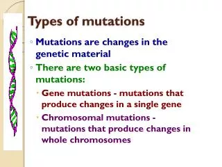
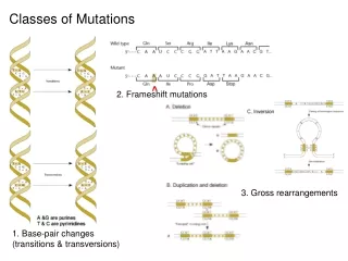
Types of Mutations Mutations can be classified in several ways - By whether they remove, alter, or add a function - By exactly how they structurally alter DNA Figure 12.4
Point Mutations A change of a single nucleotide Transition = Purine replaces purine or pyrimidine replaces pyrimidine A to G or G to A or C to T or T to C Transversion = Purine replaces pyrimidine or pyrimidine replaces purine A or G to T or C T or C to A or G Figure 12.4
Consequences of Point Mutations Missense mutation = Replaces one amino acid with another Nonsense mutation = Changes a codon for an amino acid into a stop codon - Creates truncated proteins that are often non-functional A stop codon that is changed to a coding codon lengthens the protein Figure 12.4
Splice Site Mutations Alters a site where an intron is normally removed from mRNA Can affect the phenotype if: 1) Intron is translated or exon skipped - Example: CF mutation 2) Exon is skipped - Example: Familial dysautonomia (FD) Figure 12.4
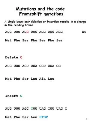
Deletions and Insertions The genetic code is read in triplets Nucleotides changes not in multiples of 3 lead to disruptions of the reading frame Cause a frameshift mutation and alter amino acids after mutation Nucleotide changes in multiples of 3 will NOT cause a frame-shift - But they can still alter the phenotype Figure 12.4
Deletions and Insertions A deletion removes genetic material - Male infertility: Tiny deletions in the Y An insertion adds genetic material - Gaucher disease: Insertion of one base A tandem duplication is an insertion of identical sequences side by side - Charcot-Marie-Tooth disease: Tandem duplication of 1.5 million bases Figure 12.4
Note: Different types of mutations can cause the same single-gene disorder - Example: Familial hypercholesterolemia Figure 12.4 Figure 12.9
Pseudogenes A DNA sequence similar to a gene but which is not translated May not even be transcribed into RNA May have evolved from original gene by duplication and acquired mutation Crossing over between a pseudogene and functional gene can disrupt gene expression Figure 12.4
Expanding Repeats Insertion of triplet repeats leads to extra amino acids - The longer proteins shut down the cells Some genes are particularly prone to expansion of repeats Number of repeats correlates with earlier onset and more severe phenotype Anticipation is the expansion of the triplet repeat with an increase in severity of phenotype with subsequent generations Figure 12.4
Myotonic Dystrophy: A Triplet Repeat Disease Figure 12.10 Figure 12.10
Copy Number Variants (CNV) Are sequences that vary in number from person to person Range in size from a few bases to millions Account for about 25% of our genome CNVs may have no effect on the phenotype or they can disrupt a gene’s function and harm health Figure 12.4
Copy Number Variants (CNV) Indeed, CNVs correlated to cholesterol level might be used to give medical advice Figure 12.4 Figure 12.11
Importance of Position The degree that a mutation alters phenotype depends on: - Where in the gene the change occurs - How it affects conformation or expression of encoded protein Examples – Hemoglobin and prions - Certain mutations exert effects while other do not Figure 12.4
Globin Mutations Table 12.5
Prion Disorders A prion disease can be inherited or acquired The prion protein exists in both normal and infectious conformations - The normal form has a central core made up of helices - In a disease-causing form, the helices open into a sheet Figure 12.4
Not All MutationsImpact Protein Function Silent mutations are mutations that do not alter the encoded amino acid Example: - A mutation from CAA to CAG alters the DNA, but the protein sequence remains unchanged - CAA and CAG both code for glutamine - These are called synonymous codons Figure 12.4
Not All MutationsImpact Protein Function A missense mutation alters the encoded amino acid to another amino acid - Creates a nonsynonymous codon Some nonsynonymous mutations are conservative; Encode a chemically similar amino acid and may not alter function The impact of a missense mutation is not predictable from protein sequence alone Figure 12.4
A conditional mutation produces a phenotype under particular conditions or environments Glucose 6-phosphate dehydrogenase enzyme responds to oxidants, chemicals that strip electrons from other molecules High levels of oxidants occur when eating fava beans or taking certain antimalarial drugs ConditionsIndividuals with G6PD mutations Low oxidants No phenotype High oxidants RBCs burst; Hemolytic anemia Figure 12.4
DNA Repair Errors in DNA replication or damage to DNA create mutations - May result in cancer Fortunately, most errors and damage are repaired Type of repair depends upon the type of damage or error Organisms vary in their ability to repair DNA Figure 12.4
Types of DNA Repair In many modern species, three types of DNA repair peruse the genetic material 1) Photoreactivation repair 2) Excision repair 3) Mismatch repair Figure 12.4
Photoreactivation Repair Enzymes called photolyases use light energy to break the extra bonds in a pyrimidine dimer Enables UV-damaged fungi to recover from exposure to sunlight Humans do not have this type of repair Figure 12.4
Excision Repair Pyrimidine dimers and surrounding bases are removed and replaced Humans have two types of excision repair Nucleotide excision repair - Replaces up to 30 bases - Corrects mutations caused by different insults Base excision repair - Replaces 1-5 bases - Specific to oxidative damage Figure 12.4
Excision Repair Figure 12.13 Figure 12.13
Figure 12.14 Mismatch Repair Enzymes detect nucleotides that do not base pair in newly replicated DNA The incorrect base is excised and replaced Proofreading is the detection of mismatches Figure 12.4