Download

1 / 17
210 likes | 588 Views
ECE 738 Project. Brain segmentation and Phase unwrapping in MRI data. JongHoon Lee. Outline. Nature of fast MRI: EPI & Field Inhomogeneity Background problem – Image Distortion Specific problems a) Brain Segmentation b) Phase Unwrapping Goal Approach Result

E N D
ECE 738 Project Brain segmentation and Phase unwrapping in MRI data JongHoon Lee
Outline • Nature of fast MRI: EPI & Field Inhomogeneity • Background problem – Image Distortion • Specific problems • a) Brain Segmentation • b) Phase Unwrapping • Goal • Approach • Result • Conclusion and future work
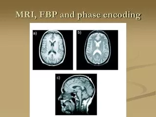
Nature of fast MRI: EPI • EPI(Echo Planar Imaging) • - Most common technique for fast MRI • Magnetic Field - B(r) • - Homogeneous w/o object : B(r) = const. • - Inhomogeneous w/ object (e.g.head: bone, brain, air,…) • > Due to magnetic susceptibility difference • > EPI is sensitive to this • Geometrical Distortion in EPI image x, y : amounts of shift (misplacement) in x, y directions in mm FOVx, FOVy : field of view in x, y direction in mm tx, ty : sampling interval in x, y direction ( 1/sampling rate)
Nature of fast MRI: EPI • Geometric Distortion Example • Significance • - Misregistration of EPI and Anatomical image • Incorrect mapping of region of interest(ROI) • Need to be corrected using Fieldmap
Specific Problems • MRI data = complex image data from Fourier transform Magnitude image Phase image Real image Imaginary image • Fieldmap = Field inhomogeneity map = TE : Echo time - Peak signal time
Specific Problems Phase Unwrapping x = I Scanner Brain Segmentation II Fieldmap Correction
Specific Problems Phase Unwrapping True phase Wrapped phase in original phase data Wrapped phase with noise Unwrapped phase: True phase Unwrapped phase: Fooled by noise
Specific Problems Phase Unwrapping Problem. 1– Imperfect segmentation Noise at boundary Erroneous unwrapping by 1D conventional Unwrapping Solution! - Unwrapping from inside to outside: Seed growing Problem. 2– Islands Erroneous unwrapping by slice by slice 2D based seed growing method Solution! – 3D volume based Unwrapping: 3D Seed growing
Specific Problems Brain Segmentation Manual Method - time cost - requirement for sufficient training Automated Method - combination of image-processing techniques thresholding | clustering | region growing edge detection | morphological | surface modeling - Seeded region growing algorithm based method - Histogram - Morphology based method - Deformable surface modeling Problem – All the segmentation method is intensity dependent May cause problem with phase map data Solution? – Segmentation usingphase map data
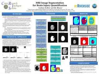
Goal SimultaneousPhase unwrapping & Segmentation of brain area by assuming smoothly varying phase in brain for Fieldmap correction of fast MRI(EPI)
Approach 3D seed growing unwrapping guided by noise-pole field. Based on papers by R. Cusack et.al. & Sofia et.al. • Generate pole field • Modify the pole field with initial thresholded mask Noise-pole field • Find purest point in the center of 3D brain data Seed • Merge or unwrap adjacent pixels Seed growing • Repeat with new, increased threshold Iteration • Stop at the final threshold Unwrapped phase map • Set nonzero brain area to ‘1’ Mask data (Segmentation) • Make fieldmap from two phase maps Geometric correction of EPI
Approach Details Generate: Noise-Pole field A’ A’ A A A A’ Pole A = A’ Phase Map Pole Field Initial Mask Noise-Pole Field • Unique point! • Noise-pole field • Computationally expensive segmentation is unnecessary
Approach Details Iterative seed growing:recursive algorithm • Implemented in Matlab • Easy to visualize/control image data • Poor to deal with recursive algorithm! ‘Out of Memory!’ Problem • Converting to ‘C’ didn’t work • Repeating ‘for loops’ in a recursive function • Increased speed and solved memory problem • ex) 20 repetition in a function reduced time 1/3 • Unwrapping from less noisy area to more noisy area
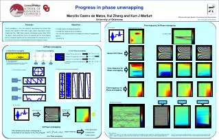
Results.1 Phase Unwrapping
Results.2 Brain Segmentation MRI intensity images Mask from new way Mask from High complexity segmentation (BET, Stephen M. Smith)
Results.3 Undistortion
Conclusion & Future Work • First work of brain segmentation using phase data • Phase only segmentation is possible research area • Reduce complexity of whole procedure of fieldmap generation • Unwrapping and segmentation are executed at the same time • Has not applied to other applications • Smoothing and threshold parameters are to be chosen carefully • Narrow areas tends to be eroded by smoothing • Implementation in other language for faster operation