Download
1 / 31
390 likes | 1.23k Views
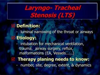
Laryngo- Tracheal Stenosis (LTS). Definition: luminal narrowing of the throat or airways Etiology: intubation for mechanical ventilation, trauma, airway surgery, reflux, malformations (EA, Vessels......) Therapy planing needs to know: number, site, degree, extent, & dynamics.

E N D
Laryngo- Tracheal Stenosis (LTS) • Definition: • luminal narrowing of the throat or airways • Etiology: • intubation for mechanical ventilation, trauma, airway surgery, reflux, malformations (EA, Vessels......) • Therapy planing needs to know: • number, site, degree, extent, & dynamics
LTT – Clin. Examination • Palpation, Percussion • Auskultation • Inspection • Indirect: Larynx only Mirror, Microlaryngoskopy • Direct: Larynx & Trachea
Imaging of LTS • Conv. X-Rays: • Chest-X, Softtissue X-Rays • Fluoroscopy • Conv. Tomography • Magnetresonance Tomography • Spiral Computertomography • 3D Reconstructions • Virtuelle Endoscopy
LTS Imaging – 3D • Multiplanare Rekonstruktionen • Minimum Intensity Projection • Shaded Surface Display • Volume Rendering • (Kombinationen)
LTS – VE „Impact“ • Study: n=19 • Patients: n=15 • Controlls: n=4 • Comparision – reporting with/without VE - 2 observers: • Axiale Schichten, MPR • Axiale Schichten, MPR und VE
LTS – VE „Impact“ E.Sorantin et al. Ped Radiol (2002) 32: 8-15
LTS Quantification – Endoscopy • High interobserverariability!!!!Jewett et al. Ann Otol Rhinol Laryngol 1999)
LTS - Quantifizierung • Visual – semi-quantitative
LTS Quantification – Spiral CT • Interobserver Variance • 3 Observer, 22 Trachealstenoseses
LTS – Quantification • 3D-Cross sectional profile: • Airway Segmentation • Extraction of the centerline: • Skeletonisation. • Orthogonal on centerline 3D cross sectional profile • Caliber change change in the cross sectional area
LTS – Quantification Vocal Chords Subgl.Space Stenosis Upper border Sternum Lower border Cricoid
LTS - ValidationPhantoms • Accuracy and Precision
Clinical Studies • Patients (n=36 24 weeks to 92a) • all invested by endoscopy and CT • Normal controls (n=18)
LTS - ValidationPhantoms • length measuremtens: 1% error
Results - Phantoms • Theoretical vs computed cross sectional profile
Conclusions • Realistic 3D reconstructions from S-CT are possible • Virtual endoscopy presents data in a familiar way for the ENT surgeon • 3D cross sectional charts: • provide quantitative information • number • site • lenght • degree
Conclusions • 3D cross sectional charts: • accurate • precise • caliber changes up to 20% in normals ROAD MAPPING ACHIEVED
Other Possibilities • Usuage of the central path: • for automated steering of a virtual camera • Volume Rendering: • adjustment of the opacity curve according the segmented airways