Download
1 / 40
400 likes | 430 Views
Learn about skeletal system function, types of joints, muscle definitions, and emergency care for fractures, dislocations, and vascular injuries in this comprehensive guide on musculoskeletal trauma.

E N D
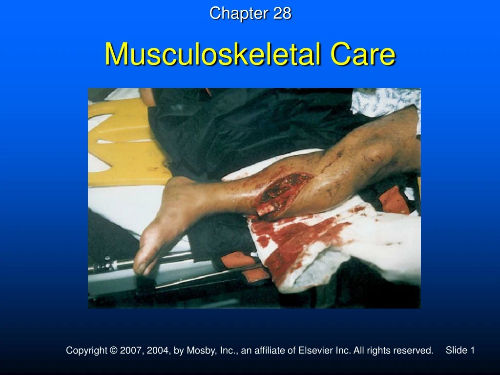
Chapter 28 Musculoskeletal Care
Case History You respond to a call for a man who fell from a roof. On arrival, you find the patient screaming in pain. You notice lacerations on both legs and bone ends protruding from one leg. He also has a deformity in the middle of his left arm.
Skeletal System Function • Gives the body shape • Protects vital organs • Provides body movement
Bones and Other Connective Tissues • 206 bones • Bones are a form of connective tissue. • Other forms of connective tissue • Cartilage • Ligaments • Tendons
Types of Muscles • Voluntary • Involuntary • Cardiac
Definitions • Fracture • A break in the continuity of bone • Sprain • Injury to ligaments, usually resulting from stretching forces • Strain • Injury to muscles or their tendons, usually from overstretching or violent contractions • Dislocation • A displacement of bones in a joint from their normal anatomic position
Average Blood Loss with Closed Fracture Site of Fracture Amount of Blood Loss Radius or ulna 250-500 ml Humerus 500-750 ml Pelvis 1500-3000 ml Femur 1000-2000 ml Tibia and fibula 500-1000 ml
Signs and Symptoms • Pain and tenderness • Deformity or angulation • Swelling and discoloration • Loss of use • Grating or crepitus • Exposed bone • Joint locked into position or dislocation • Bleeding
Vascular Injuries • Vessels can be compressed or torn. • Dislocations are at high risk for vessel compression or injury. • Loss of blood flow to extremity
Vascular Injuries –Signs and Symptoms • Loss of distal pulses • Pale, cool skin • Delayed or absent capillary refill • Pain • Numbness • Tingling or prickling • Sensory loss • Paralysis
Vascular Injuries Memory Device –The “5 Ps” • Pain • Pallor • Pulselessness • Paresthesia (tingling) • Paralysis
Peripheral Nerve Injury • Injuries can cause compression or complete tearing of nerve. • Violent forces can cause direct damage. • Nerve injury occurs commonly in joints.
Signs and Symptoms • Pain and tenderness • Deformity or angulation • Loss of use • Grating • Swelling • Bruising (discoloration) • Exposed bone ends • Joint locked into position
Emergency Medical Care • Use personal protection measures. • Administer oxygen. • After life threats have been controlled, splint injuries in preparation for transport. • Apply a cold pack. • Elevate the extremity.
Splinting • Reasons • Prevent motion of bone fragments • Minimize the following complications: • Damage to muscles, nerves, or blood vessels • Conversion of a closed fracture to an open fracture • Restriction of blood flow • Excessive bleeding • Increased pain • Paralysis of extremities due to a damaged spine • Injury to viscera
General Rules of Splinting • Assess pulse, motor function, and sensation. • Before and after splint application • Record findings. • Immobilize the joint above and below the injury.
General Rules of Splinting • Remove or cut away clothing. • Cover open wounds with a sterile dressing. • Severe deformity, cyanotic distal extremity, or lack of pulses • Align with gentle traction before splinting.
General Rules of Splinting • Do not intentionally replace the protruding bones. • Pad each splint to prevent pressure and discomfort to the patient. • Splint the patient before moving when feasible and no life threats.
General Rules of Splinting • When in doubt, splint the injury. • If patient has signs of shock • Align in normal anatomic position • Transport on backboard
Equipment • Rigid splints • Traction splints • Pneumatic splints (air, vacuum) • Improvised splints, pillow • Pneumatic antishock garment (as a splint)
Hazards of Improper Splinting • Compression of nerves, tissues, and blood vessels • Delay in transport of patient with life-threatening injury
Hazards of Improper Splinting • Splint applied too tight • Aggravation of bone or joint injury • Cause or aggravate tissue, nerve, vessel, or muscle damage • From excessive bone or joint movement
Long Bone Splinting • Use personal protection measures. • Apply manual stabilization. • Assess pulse, motor and sensory function before and after splinting. • Severe deformity, cyanotic distal extremity, or lack of pulses • Align with gentle traction before splinting. • Apply splint. • Immobilize hand/foot in position of function.
Traction Splinting –Indications • Mid-thigh injury • No joint or lower leg injury
Contraindications • Injury close to the knee • Injury to the knee • Injury to the hip • Injured pelvis
Contraindications • Partial amputation or avulsion with bone separation • Distal limb is connected only by marginal tissue. • Traction would risk separation. • Lower leg or ankle injury
Traction Splinting – Procedure • Use personal protection measures. • Assess pulse, motor function, and sensation before and after splinting. • Perform manual stabilization of the injured leg. • Apply splint.
Traction Splinting – Procedure • Reevaluate proximal/distal securing devices. • Reassess pulse, motor and sensory function. • Secure torso to the long board. • Secure splint to the long board to prevent movement of splint.
Clavicle Shoulder Upper arm Elbow Forearm/wrist Hand Pelvis Sling and swathe Sling and swathe Sling and swathe with splint Rigid splint/sling Sling and swathe with splint Rigid splint and sling Long spine board/PASG Specific Splinting Techniques —Upper Body
Dislocated hip Hip injury Femur Knee Lower leg Ankle and foot Anatomic/spine board Anatomic/spine board Traction splint/PASG Rigid splint Rigid splint Pillow splint Specific Splinting Techniques —Lower Body