Immune Systems: Innate and Acquired Defense Mechanisms
630 likes | 691 Views
Explore the innate and acquired immunity mechanisms in animals, including external barriers, phagocytic cells, and lymphocytes. Learn how these defenses recognize and respond to pathogens to protect the body from infections.

Immune Systems: Innate and Acquired Defense Mechanisms
E N D
Presentation Transcript
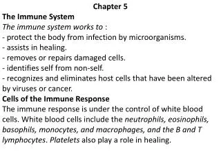
Reconnaissance, Recognition, and Response • An animal must defend itself • From the many dangerous pathogens it may encounter in the environment • Two major kinds of defense have evolved that counter these threats • Innate immunity and acquired immunity
Figure 43.1 3m • Innate immunity • Present before any exposure to pathogens and is effective from the time of birth • Involves nonspecific responses to pathogens
Acquired immunity, also called adaptive immunity • Develops only after exposure to inducing agents such as microbes, toxins, or other foreign substances • Involves a very specific response to pathogens
INNATE IMMUNITY Rapid responses to a broad range of microbes ACQUIRED IMMUNITY Slower responses to specific microbes External defenses Internal defenses Skin Phagocytic cells Humoral response (antibodies) Mucous membranes Antimicrobial proteins Secretions Inflammatory response Invading microbes (pathogens) Cell-mediated response (cytotoxic lymphocytes) Natural killer cells Figure 43.2 • A summary of innate and acquired immunity
Innate immunity provides broad defenses against infection • Pathogen that successfully breaks through an animal’s external defenses • encounters innate mechanisms that impede its attack
External Defenses • Intact skin and mucous membranes • Physical barriers • Mucus • Traps microbes and other particles
10m Figure 43.3 • Ciliated epithelial cells (trachea) • Sweep mucus and any entrapped microbes upward, preventing the microbes from entering the lungs
Secretions of the skin and mucous membranes • Provide an environment that is often hostile to microbes • Secretions from the skin • pH of 3 and 5, prevents colonization of many microbes • Also lysozyme, destroys bacteria
Internal Cellular and Chemical Defenses • Phagocytes, (white blood cells) • Ingest invading microorganisms • Initiate the inflammatory response
Pseudopodia surround microbes. 1 Microbes Microbes are engulfed into cell. 2 MACROPHAGE Vacuole containing microbes forms. 3 Vacuole Lysosome containing enzymes Vacuole and lysosome fuse. 4 Toxic compounds and lysosomal enzymes destroy microbes. 5 Microbial debris is released by exocytosis. 6 Phagocytic Cells • Attach to their prey via surface receptors and engulf them Figure 43.4
Macrophages( a type of phagocyte) • migrate through the body • Also in organs of the lymphatic system
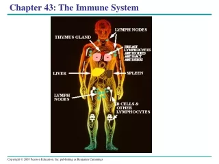
Interstitial fluid bathing the tissues, along with the white blood cells in it, continually enters lymphatic capillaries. 1 Lymphatic capillary Interstitial fluid Fluid inside the lymphatic capillaries, called lymph, flows through lymphatic vessels throughout the body. 2 Adenoid Tonsil Lymphatic vessels return lymph to theblood via two largeducts that drain intoveins near the shoulders. 4 Lymph nodes Blood capillary Spleen Lymphatic vessel Tissue cells Peyer’s patches (small intestine) Within lymph nodes, microbes and foreign particles present in the circulating lymph encounter macro- phages, dendritic cells, and lymphocytes, which carry out various defensive actions. 3 Appendix Lymphatic vessels Masses of lymphocytes and macrophages Lymph node Figure 43.5 • The lymphatic system
Antimicrobial Proteins • Attack microbes directly or impede their reproduction
e.g. the complement system • Cause lysis of invading cells and help trigger inflammation • Interferons • Defense against viruses
Inflammatory Response • Histamine and other chemicals released from injured cells • changes in blood vessels that allow more fluid, more phagocytes, and antimicrobial proteins to enter the tissues
Blood clot Pin Pathogen Macrophage Blood clotting elements Chemical signals Phagocytic cells Phagocytosis Capillary Red blood cell Fluid, antimicrobial proteins, and clotting elements move from the blood to the site. Clotting begins. 1 Chemical signals released by activated macrophages and mast cells at the injury site cause nearby capillaries to widen and become more permeable. Neutrophils and macrophages phagocytose pathogens and cell debris at the site, and the tissue heals. Chemokines released by various kinds of cells attract more phagocytic cells from the blood to the injury site. 4 2 3 • Major events in the local inflammatory response Figure 43.6
Natural Killer (NK)Cells • Patrol the body and attack virus-infected body cells and cancer cells • Trigger apoptosis (cell death)
Invertebrate Immune Mechanisms • Similar to vertebrate innate response
Acquired immunity • Lymphocytes provide specific defenses against infection • Body’s second major kind of defense
Antigen- binding sites Epitopes (antigenic determinants) Antibody A Antigen Antibody B Antibody C • Antigen (foreign molecule) • Specifically recognized by lymphocytes and elicits a response from them • Lymphocyte recognizes and binds to just a small portion of the antigen called an epitope Figure 43.7
Antigen Recognition by Lymphocytes • 2 main types of lymphocytes • B lymphocytes (B cells) and T lymphocytes (T cells), circulate through the blood
Antigen- binding site Antigen- binding site Disulfide bridge V V V V Variable regions Light chain C C Constant regions C C Transmembrane region Plasma membrane Heavy chains B cell Cytoplasm of B cell (a) A B cell receptor consists of two identical heavy chains and two identical light chains linked by several disulfide bridges. Figure 43.8a B Cell Receptors for Antigens • Bind to specific antigens • Called antibodies or immunoglobulins, 4 chains, 2 heavy, 2 light
Antigen- Binding site Variable regions Constant regions Transmembrane region b chain Plasma membrane a chain Disulfide bridge T cell Cytoplasm of T cell (b) • A T cell receptor consists of one • chain and one b chain linked by a disulfide bridge. T Cell Receptors • 2 different polypeptide chains V V C C Figure 43.8b
T cells bind to small fragments of antigens bound to cell-surface proteins called MHC molecules
Infected cells produce MHC molecules which bind to antigen fragments which are presented on surface of cell • T cells then detect the antigen fragment displayed on the cell’s surface
1 Infected cell A fragment of foreign protein (antigen) inside the cell associates with an MHC molecule and is transported to the cell surface. 1 Antigen fragment 2 Class I MHC molecule 2 The combination of MHC molecule and antigen is recognized by a T cell, alerting it to the infection. T cell receptor (a) Cytotoxic T cell Figure 43.9a • Class I MHC molecules, found on almost all nucleated cells of the body • Display peptide antigens to cytotoxic T cells
1 Antigen- presenting cell Microbe A fragment of foreign protein (antigen) inside the cell associates with an MHC molecule and is transported to the cell surface. 1 Antigen fragment 2 2 Class II MHC molecule The combination of MHC molecule and antigen is recognized by a T cell, alerting it to the infection. T cell receptor Helper T cell (b) • Class II MHC molecules, located mainly on dendritic cells, macrophages, and B cells • Display antigens to helper T cells Figure 43.9b
Lymphocyte Development • Arise from stem cells in the bone marrow
Bone marrow Lymphoid stem cell Thymus T cell B cell Blood, lymph, and lymphoid tissues (lymph nodes, spleen, and others) Figure 43.10 • Newly formed lymphocytes are all alike • But they later develop into B cells or T cells, depending on where they continue their maturation
V4–V39 3 2 4 1 DNA of undifferentiated B cell V40 J1 J2 J3 J4 J5 V2 V3 Intron V1 C Deletion of DNA between a V segmentand J segment and joining of the segments DNA of differentiated B cell V3 V1 V2 J5 Intron C Transcription of resulting permanently rearranged,functional gene V3 J5 C pre-mRNA Intron RNA processing (removal of intron; addition of capand poly (A) tail) V3 J5 C Poly (A) mRNA Cap Translation V C Light-chain polypeptide B cell receptor Variable region Constant region B cell Figure 43.11 • Immunoglobulin gene rearrangement
Antigen molecules bind to the antigen receptors of only one of the three B cells shown. Antigen molecules B cells that differ in antigen specificity Antigen receptor The selected B cell proliferates, forming a clone of identical cells bearing receptors for the selecting antigen. Some proliferating cells develop into short-lived plasma cells that secrete antibodies specific for the antigen. Some proliferating cells develop into long-lived memory cells that can respond rapidly upon subsequent exposure to the same antigen. Antibody molecules Clone of memory cells Clone of plasma cells Figure 43.12 Clonal selection of B cells • Generates a clone of short-lived activated effector cells and a clone of long-lived memory cells
1 Secondary response to anti- gen A produces antibodies to A; primary response to anti- gen B produces antibodies to B Day 1: First exposure to antigen A 4 Primary response to antigen A produces anti- bodies to A 2 Day 28: Second exposure to antigen A; first exposure to antigen B 3 104 103 Antibody concentration (arbitrary units) 102 Antibodies to A Antibodies to B 101 100 35 28 21 42 49 56 0 14 7 Figure 43.13 Time (days) • In the secondary immune response • Memory cells facilitate a faster, more efficient response
Humoral immune response activation and clonal selection of B cells, production of antibodies • Cell-mediated immune responseactivation and clonal selection of cytotoxicT cells
Cell-mediated immune response Humoral immune response First exposure to antigen Antigens displayedby infected cells Antigens engulfed and displayed by dendritic cells Intact antigens Activate Activate Activate Secreted cytokines activate B cell HelperT cell CytotoxicT cell Gives rise to Gives rise to Gives rise to Active and memory helperT cells Memory cytotoxicT cells Active cytotoxicT cells MemoryB cells Plasmacells Defend against infected cells, cancer cells, and transplanted tissues Secrete antibodies that defend againstpathogens and toxins in extracellular fluid Figure 43.14 • Major participants in the acquired immune response
Helper T Cells: A Response to Nearly All Antigens • Produce CD 4, a surface protein • enhances binding to class II MHC molecule–antigen complexes on antigen-presenting cells • Activation of the helper T cell then occurs
After a dendritic cell engulfs and degrades a bacterium, it displays bacterial antigen fragments (peptides) complexed with a class II MHC molecule on the cell surface. A specific helper T cell binds to the displayed complex via its TCR with the aid of CD4. This interaction promotes secretion of cytokines by the dendritic cell. Cytotoxic T cell Dendritic cell Peptide antigen Helper T cell Cell-mediated immunity (attack on infected cells) Class II MHC molecule Bacterium TCR Humoral immunity (secretion of antibodies by plasma cells) CD4 Dendritic cell Cytokines B cell Proliferation of the T cell, stimulated by cytokines from both the dendritic cell and the T cell itself, gives rise to a clone of activated helper T cells (not shown), all with receptors for the same MHC–antigen complex. The cells in this clone secrete other cytokines that help activate B cells and cytotoxic T cells. 3 1 2 3 2 1 Figure 43.15 • The role of helper T cells in acquired immunity
Cytotoxic T cells • Bind to infected cells, cancer cells, and transplanted tissues
The granzymes initiate apoptosis within the target cells, leading to fragmentation of the nucleus, release of small apoptotic bodies, and eventual cell death. The released cytotoxic T cell can attack other target cells. The activated T cell releases perforin molecules, which form pores in the target cell membrane, and proteolytic enzymes (granzymes), which enter the target cell by endocytosis. A specific cytotoxic T cell binds to a class I MHC–antigen complex on a target cell via its TCR with the aid of CD8. This interaction, along with cytokines from helper T cells, leads to the activation of the cytotoxic cell. Cytotoxic T cell Released cytotoxic T cell Perforin Cancer cell Granzymes Apoptotic target cell TCR CD8 Class I MHC molecule Pore 1 3 2 3 1 2 Target cell Peptide antigen Cytotoxic T cell Figure 43.16 • Activated cytotoxic T cell secretes proteins that destroy the infected target cell
B Cells • Respond to Extracellular Pathogens
Clonal selection of B cells • Generates antibody-secreting plasma cells, the effector cells of humoral immunity
A B cell that has taken up and degraded the same bacterium displays class II MHC–peptide antigen complexes. An activated helper T cell bearing receptors specific for the displayed antigen binds to the B cell. This interaction, with the aid of cytokines from the T cell, activates the B cell. After a macrophage engulfs and degrades a bacterium, it displays a peptide antigen complexed with a class II MHC molecule. A helper T cell that recognizes the displayed complex is activated with the aid of cytokines secreted from the macrophage, forming a clone of activated helper T cells (not shown). The activated B cell proliferates and differentiates into memory B cells and antibody-secreting plasma cells. The secreted antibodies are specific for the same bacterial antigen that initiated the response. Bacterium Macrophage Peptide antigen Class II MHCmolecule CD4 TCR B cell Secreted antibodymolecules Clone of plasma cells Endoplasmicreticulum of plasma cell Helper T cell Activated helper T cell Cytokines 3 2 3 1 1 2 Clone of memoryB cells Figure 43.17
IgM (pentamer) First Ig class produced after initial exposure to antigen; then its concentration in the blood declines Promotes neutralization and agglutination of antigens; very effective in complement activation (see Figure 43.19) J chain IgG (monomer) Most abundant Ig class in blood; also present in tissue fluids Only Ig class that crosses placenta, thus conferring passive immunity on fetus Promotes opsonization, neutralization, and agglutination of antigens; less effective in complement activation than IgM (see Figure 43.19) IgA (dimer) Present in secretions such as tears, saliva, mucus, and breast milk Provides localized defense of mucous membranes by agglutination and neutralization of antigens (see Figure 43.19) J chain Secretory component Presence in breast milk confers passive immunity on nursing infant IgE (monomer) Triggers release from mast cells and basophils of histamine and other chemicals that cause allergic reactions (see Figure 43.20) IgD (monomer) Present primarily on surface of naive B cells that have not been exposed to antigens Acts as antigen receptor in antigen-stimulated proliferation and differentiation of B cells (clonal selection) Transmembrane region Figure 43.18 5 classes of antibodies (immunoglobulins)
Binding of antibodies to antigens inactivates antigens by Viral neutralization (blocks binding to host) and opsonization (increases phagocytosis) Agglutination of antigen-bearing particles, such as microbes Activation of complement system and pore formation Precipitation of soluble antigens Complement proteins Bacteria Virus MAC Pore Soluble antigens Bacterium Foreign cell Leads to Enhances Cell lysis Phagocytosis Figure 43.19 Macrophage • Antibody-mediated mechanisms of antigen disposal
Active Immunization • Develops naturally in response to an infection • Can also develop following immunization, also called vaccination
In immunization • A nonpathogenic form of a microbe or part of a microbe elicits an immune response to an immunological memory for that microbe
Passive immunity • Provides immediate, short-term protection • Mother fetus
Ability to distinguish self from nonself • Wage war against cells from other individuals • Transplanted tissues usually destroyed by the recipient’s immune system
Blood Groups and Transfusions • Certain antigens on red blood cells • Determine whether a person has type A, B, AB, or O blood
Antibodies to nonself blood types • Already exist in the body • Transfusion with incompatible blood • Leads to destruction of the transfused cells