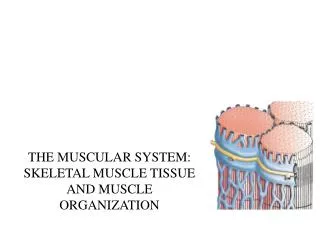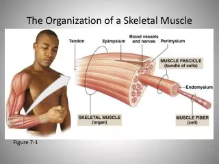
Skeletal Organization
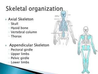
The human skeleton is divided into two main parts: the Axial Skeleton and the Appendicular Skeleton. The Axial Skeleton consists of the skull, vertebral column, and thoracic cage, whereas the Appendicular Skeleton includes the pectoral girdle, upper limbs, pelvic girdle, and lower limbs. This guide provides detailed insights into the structures of the skeleton, including major bones and terminology used to describe skeletal formations. Understanding these components is crucial for studying human anatomy and physiology effectively.


Skeletal Organization
E N D
Presentation Transcript
Skeletal Organization 7.5 p. 205 – p. 259
Basics • 206 bones in human body • Made up of 2 parts: • Axial Skeleton • Appendicular Skeleton • http://www.getbodysmart.com/ap/skeletalsystem/skeleton/introduction/tutorial.html
Axial Skeleton • Skull • Cranium • Face • Hyoid Bone • Vertebral Column • Thoracic Cage • Ribs • Sternum
Appendicular Skeleton • Pectoral girdle • Upper limbs • Pelvic girdle • Lower limbs
Terms Used to Describe Skeletal Structures (Table 7.4 p. 208) • Meatus • Process • Ramus • Sinus • Spine • Suture • Trochanter • Tubercle • Tuberosity • Condyle • Crest • Epicondyle • Facet • Fissure • Fontanel • Foramen • Fossa • Head • Linea
Skull • Cranial bones • Facial bones
Cranial Bones • Frontal bone • Parietal Bone • Temporal Bones • Occipital Bone • Sphenoid Bone • Ethmoid Bone
Cranial Bones • Frontal Bone • Forehead • Bone under eyebrows • Superior portion of eye orbits • Parietal Bones • Most superior and lateral walls of cranium • Sagittal suture at midline • Coronal suture where they meet the frontal bone
Cranial Bones • Temporal Bones: inferior to the parietal bones • Joined at squamous sutures • External acoustic meatus • Styloid process • Zygomatic process • Mastoid process • Jugular foramen • Internal acoustic meatus • Carotid
Cranial Bones • Occipital Bone • Joined to parietal bones anteriorly by the lambdoid suture • Foramen magnum • Surrounds lower part or brain, allows spinal cord to connect to brain • Occipital condyles
Cranial Bones • Sphenoid Bone • Spans width of the skull • Forms floor of cranial cavity • Stella turcica • Foramen ovale • Optic canal • Superior orbital fissure • Sphenoid sinuses
Cranial Bones • Ethmoid Bones • Anterior to sphenoid bone • Forms roof of nasal cavity • Cristagalli • Cribriform plates • Superior and middle nasal conchae
Cranial Bones Link • http://www.getbodysmart.com/ap/skeletalsystem/skeleton/axial/skull/quizzes/menu/menu.html
Facial Bones • Maxillae • Palantine Bones • Zygomatic Bones • Lacrimal Bones • Nasal Bones • Vomer Bone • Inferior Nasal Conchae • Mandible
Facial Bones • Maxillae (maxillary bones) • Upper jaw • Keystone bones – all other bones join with the maxillae bones • Upper teeth in alveolar margin • Palantine processes – anterior portion of hard palate • Paranasal sinuses • Palantine Bones: form posterior portion of hard palate • Failure to develop forms a cleft palate
Facial Bones • Zygomatic Bones • Cheek bones • Eye sockets • Lacrimal Bones • Medial portion of each eye socket • Groove to serve as passage way for tears • Nasal bones • Small bones forming bridge of the nose
Facial Bones • Vomer Bone • Nasal septum • Inferior Nasal Conchae • Thin, curved bones from the lateral walls of the nasal cavity • Mandible • Lower jaw • Body • Rami • Lower teeth in alveoli (alveolar margin)
Hyoid Bone • Only bone of the body that does not directly articulate with another bone • Suspended in neck approximately 2cm above the larynx • Movement of tongue • Attachment for neck muscles
Skull Development • Use your books and computers to summarize the development of the skull. Focus in fetal development through adolescence. • Bullet points or paragraph • Be prepared to present all or a portion to the class.
Vertebral Column • Regions: • Cervical • Thoracic • Lumbar • Sacral • Shapes vary • Intervertebral discs • Separate and cushion vertebrae
Vertebral Column Terminology • Body / centrum • Vertebral arch • Vertebral foramen • Pedicles Transverse processes Spinous processes Superior and inferior articular facets Anterior/posterior longitudinal ligaments
Cervical Vertebrae • C1 – C7 • Neck region of spine • First two vertebrae: atlas and axis
Cervical Vertebrae • Smallest vertebrae • Transverse process contains foramina for the vertebral arteries to pass through
Thoracic Vertebrae • T1 – T12 • Larger than cervical vertebrae • Connect with ribs
Lumbar Vertebrae • L1 – L5 • Sturdiest vertebrae • Massive bodies, short spinous processes
Sacrum • Fusion of 5 vertebrae • Superiorly connects with L5 • inferiorly connects with coccyx • Alae: articulate with hips • Median sacral crest • Posterior sacral foramina • Sacral canal • Sacral haitus
Coccyx • Fusion of three irregularly shaped vertebrae • “tailbone”
Parts of Vertebrae • http://www.purposegames.com/game/fb1ba7f9d8
Identify the pictures using your notes. • Cervical, thoracic, lumbar, axis, atlas
Thoracic Cage • Ribs • Sternum • Costal Cartilage
Ribs • 12 pairs of ribs (24 total) • True Ribs • First 7 pairs of ribs • Vertebrosternal ribs • False ribs: • Next 5 pairs of ribs • Do not reach sternum directly • Last 2 pairs of the false ribs are sometimes called floating ribs
Sternum • Midline of anterior portion of thoracic cage • 3 parts: • Manubrium • Body • Xiphoid process
Pectoral Girdle • Shoulder girdle • Clavicles • Scapulae (scapula) • Support of upper limb and muscle attachment
Clavicle • Slender, rod like bones • Elongated S-shape • Base of the neck • Run from the manubrium to the scapulae
Scapulae • Broad, triangular in shape • Either side of the upper back • Spine • Acromion process and coracoid process form the top of the shoulder • Glenoid cavity – articulates with the head of the humerus
Upper Limb • Humerus • Radius • Ulna • Hand • Carpals • Metacarpals • Phalanges
Humerus • Scapula elbow • Head fits into the glenoid cavity of the scapula • Greater tubercle • Lesser tubercle • Both provide muscle attachment
Radius • Thumb side of the forearm • Shorter than the ulna • Head articulates with the ulna (radial notch) and the humerus
Ulna • Longer than the radius • Articulates with the humerus (trochlear notch)
Hands • Wrist • 8 small carpals in two rows • All together called carpus • Palm • 5 bones, in line with each finger • Fingers • Phalanges • 2 bones in thumb • 3 bones in fingers
Pelvic Girdle • Hip bones • Ilium • Ischium • Pubis • Symphysis pubis • Male pelvis is more slender than the female pelvis • Female ilium is wider
Lower Limb • Femur • Patella • Tibia • Fibula • Foot: • Tarsals • Metatarsals • Phalanges
Femur • Longest bone in the body • Connects hip to knee • Greater trochanter, lesser trochanter • Lateral and medial chondyle articulate with tibia
Patella • Kneecap • Covers joint between femur and tibia
Fibula and Tibia • Fibula • Bears no body weight • Lateral portion of the leg • Protrudes from ankle • Tibia: shin bone • Larger of the two • Articulates at the knee with the femur • Medial portion of lower leg
Foot • Tarsals (form the tarsus) • Talus: forms medial portion of ankle • Calcaneus: heel bone • Metatarsals: 5 bones • Form foot • Phalanges • Toes • Big toe (2 bones) • other 4 toes (3 bones)