Download
1 / 35
350 likes | 801 Views
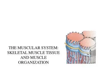
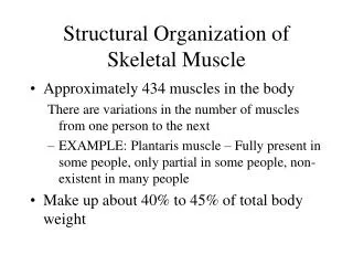
Structural Organization of Skeletal Muscle. Approximately 434 muscles in the body There are variations in the number of muscles from one person to the next EXAMPLE: Plantaris muscle – Fully present in some people, only partial in some people, non-existent in many people

E N D
Structural Organization of Skeletal Muscle Approximately 434 muscles in the body There are variations in the number of muscles from one person to the next EXAMPLE: Plantaris muscle – Fully present in some people, only partial in some people, non-existent in many people Make up about 40% to 45% of total body weight
Knee Flexors Plantaris
Palmaris Longus Forearm Muscles – Palmar View
Muscle Cells • Called muscle fibers because of their threadlike shape • Sarcolemma – Membrane surrounding the muscle cells
Muscle Cells (continued) • Sarcoplasm – Cytoplasm of muscle cells Contains: • Multiple nuclei for each cell • Mitochondria • Myofibrils or Myofilaments – Threadlike structures containing the contractile proteins Actin and Myosin • Sarcoplasmic Reticulum and Transverse Tubules – Pathways that allow the passage of the electrochemical mediators of muscle activation.
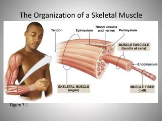
Muscle Cells (continued) • Endomysium – Connective tissue surrounding the sarcolemma • Fascicles – Parallel bundles of muscle fibers • Perimysium – Connective tissue sheath surrounding fascicles • Epimysium – Connective tissue sheath surrounding bundles of fascicles • The epimysium is continuous with the muscle tendons • Fascia – White, fibrous connective tissue surrounding entire muscles and making up the structure of the perimysium and epimysium.
Muscle Fiber Growth • Length and diameter increase from birth to adulthood through the process of natural maturation • Diameter can be increased through resistance training (hypertrophy)
Muscle Fiber Growth (continued) • The number of fibers is genetically determined and varies from person to person • Hyperplasia – An increase in the number of muscle fibers as a result of resistance training • MAY occur in some individuals • NOT a proven phenomenon • Seems to be related to the type of stress and/or damage applied to the muscle
Motor Units • Motor Neurons – Nerve cells that transmit a stimulus to muscle tissue. • Motor Unit – A functional group of muscle fibers containing a single motor neuron and the muscle fibers innervated by it. • Axon – A threadlike process forming the central core of a motor neuron.
Motor Units (continued) • Each axon will split up to contact individual muscle fibers • Motor End Plate (also called the neuromuscular junction or myoneural junction) • The area of contact between a motor neuron and a muscle cell • In humans there is one motor end plate per skeletal muscle fiber.
Motor Units (continued) • A single neuron may activate from less than 100 to nearly 2000 fibers • Fine movements (eyes, fingers, etc) are produced by motor units with a small number of fibers • Gross movements (large applications of force as in the gastrocnemius) are usually produced by motor units with large numbers of fibers
Motor Units (continued) • Twitch Type Cells • Develop tension as a result of a single stimulus • Make up most skeletal muscle units • Reach a peak value of tension very rapidly (less than 100 msec) and then lose tension immediately • Summation – Activation of a muscle fiber by rapid successive impulses until the maximum tension for that fiber is reached • Tetanus – A condition in which peak fiber tension is reached but not released
Motor Units (continued) • Tonic Type Cells • Motor units which require more than a single stimulus to develop tension • Found in the oculomotor apparatus (controls eye movement)
Muscle Spindles • Sensory receptors spread throughout the muscle fibers • Parallel to the muscle fibers • Composed of about 3-10 small muscle fibers encased in a sheath of connective tissue • Designed to respond to changes an increase in muscle length (stretching)
Muscle Spindles (continued) • Two Types. • Primary Spindles – Respond to amount of elongation and speed of elongation (dynamic response). • Secondary Spindles – Respond only to the amount of elongation (static response). The dynamic response is much stronger than the static response. Therefore a slow stretch of the muscle doesn’t activate the muscle spindle response.
Stretch Reflex • Also called the myotatic reflex • Activated by the muscle spindles in muscles undergoing rapid stretching • Produces tension in the muscles being stretched • May serve a protective function to prevent joint damage
Reciprocal Inhibition • Activated by the muscle spindles in muscles undergoing rapid stretching • Inhibits or prevents the development of tension in muscles that are antagonists to the muscles being stretched
Types of Skeletal Muscle Fibers • Classification is based on the time it takes to reach maximum tension. • Basic Categories • Slow-Twitch (ST) • Fast-Twitch (FT) It takes FT fibers about 1/7 as long to reach maximum tension as ST fibers.
Tension Developed Over Time in Muscle Fibers text page 152
Types of Skeletal Muscle Fibers (continued) There is a wide range of twitch times to reach maximum tension within both categories. FT fibers are larger in diameter than ST fibers. FT fibers usually fatigue more rapidly than ST fibers.
Types of Skeletal Muscle Fibers (continued) Intact FT and ST fibers will generate about the same peak isometric force per cross sectional area of muscle. Individuals with a higher percent of FT fibers are able to generate more torque and power during movement than people with a higher percent of ST fibers.
Types of Skeletal Muscle Fibers (continued) There are FT fibers that exhibit endurance characteristics similar to ST fibers. For this reason FT fibers can be broken down into two sub-categories, resulting in a total of three muscle fiber types.
Types of Skeletal Muscle Fibers (continued) • Type I • Slow-Twitch Oxidative (SO) • Related to lower intensity endurance activities (long distance running – marathon, etc) • Type IIA • Fast-Twitch Oxidative Glycolytic (FOG) • Related to higher intensity activities requiring endurance (middle distance running – 1500 m, 5000 m, etc)
Types of Skeletal Muscle Fibers (continued) • Type IIB • Fast-Twitch Glycolytic (FG) • Related to high intensity short duration activities (sprint races – 60 m, 200 m, etc) All fibers in a single motor unit are the same type.
Types of Skeletal Muscle Fibers (continued) Most skeletal muscles contain both ST and FT muscle fibers with the relative amounts varying between muscles. EXAMPLE: In the calf, the soleus is mainly used for maintaining basic balance and posture so it primarily contains ST fibers. The gastrocnemius is used more for quick, power related movements so it tends to have a higher percentage of FT fibers. In the general population most people have about the same percent of ST and FT fibers.
Types of Skeletal Muscle Fibers (continued) World class endurance athletes have been shown to have a very high percent of ST fibers while elite athletes in speed and power events tend to have a high percent of FT fibers.
Types of Skeletal Muscle Fibers (continued) • There seems to be a decrease in the number of FT fibers with age. • Infants and young children seem to have a lower percentage of Type IIB fibers than adults. • Significantly lower proportions of Type IIB fibers are found in obese versus non-obese individuals.