Download
1 / 13
130 likes | 272 Views
Motion estimation with tagged ultrasound images. *H. Liebgott – Adeline Bernard – Sébastien Salles. Introduction of the platform scanners/ probes / phantoms. Tagging: a way to facilitate motion estimation. Conventional MRI sequence. Tagged MRI sequence.

E N D
Motion estimation with tagged ultrasound images *H. Liebgott – Adeline Bernard – Sébastien Salles
Introduction of the platform scanners/ probes / phantoms
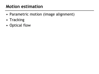
Tagging: a way to facilitate motion estimation Conventional MRI sequence Tagged MRI sequence
US Tagging or Transverse oscillations 49.5 Depth [mm] 50 50.5 -1 -0.5 0 0.5 1 1 Amplitude 0.5 Amplitude 0 -0.5 -1 -1 -0.5 0 0.5 1 -1 Lateral position [mm] x z Conventional *PSF Axial motion estimation OK Transvers motion estimation more difficult *PSF = Point spreadfunction, image of a single scatterrer
US Tagging or Transverse oscillations 49.5 Depth [mm] 50 50.5 -1 -0.5 0 0.5 1 1 Amplitude 0.5 Amplitude 0 -0.5 -1 -1 -0.5 0 0.5 1 -1 Lateral position [mm] 49.5 Depth [mm] 50 50.5 x 1 -1 -0.5 0 0.5 1 Amplitude 0.5 z Amplitude 0 -0.5 -1 -1 -0.5 0 0.5 1 Lateral position [mm] Conventional *PSF ???Tagged PSF ??? *PSF = Point spreadfunction, image of a single scatterrer
TO image formation principle US Taggingiscreatedusingspecificbeamforming*strategies It canbeseen as the interferencesbetweentwo sources *Beamforming = combination of the signalsreceived by the probe’selements
US-Taggingimages on the Ula-Op • Images • Transverse motion estimation on a phantom • 2D motion estimation of the carotidartery
US-Tagging and cardiacimaging Simulations FFT 2D Conventionnelle US-Tagging
in vivo Conventionnel US-Tagging
Conclusion • « US tagging » • Facilitate 2D motion estimation • Realistic simulations • faisabilité in vitro and in vivo • Perspectives • Validatequantitatively the in vivo part • Ultrafast US Tagging • 3D US Tagging