Brainstem
600 likes | 803 Views
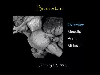
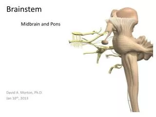
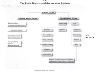
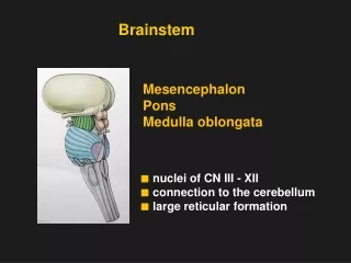
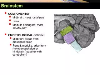
Brainstem. COMPONENTS: Midbrain: most rostal part Pons Medulla oblongata: most caudal part EMBRYOLOGICAL ORIGIN: Midbrain : arises from mesencephalon Pons & medulla : arise from rhombencephalon or hindbrain (together with cerebellum). Brainstem. COMPONENTS:

Brainstem
E N D
Presentation Transcript
Brainstem • COMPONENTS: • Midbrain: most rostal part • Pons • Medulla oblongata: most caudal part • EMBRYOLOGICAL ORIGIN: • Midbrain: arises from mesencephalon • Pons & medulla: arise from rhombencephalon or hindbrain (together with cerebellum)
Brainstem • COMPONENTS: • Midbrain: most rostal part • Pons • Medulla oblongata: most caudal part • EMBRYOLOGICAL ORIGIN: • Midbrain: arises from mesencephalon • Pons & medulla: arise from rhombencephalon or hindbrain (together with cerebellum)
Brainstem • COMPONENTS: • Midbrain: most rostal part • Pons • Medulla oblongata: most caudal part • EMBRYOLOGICAL ORIGIN: • Midbrain: arises from mesencephalon • Pons & medulla: arise from rhombencephalon or hindbrain (together with cerebellum) Anterior view
Brainstem • COMPONENTS: • Midbrain: most rostal part • Pons • Medulla oblongata: most caudal part • EMBRYOLOGICAL ORIGIN: • Midbrain: arises from mesencephalon • Pons & medulla: arise from rhombencephalon or hindbrain (together with cerebellum) Posterior view
Brainstem • SITE: • It lies on the basilar part of occipital bone (clivus). • The midbrain is continuous rostrally with diencephalon of forebrain. • The pons is continous rostrally with midbrain & caudally with medulla. • The medulla is continuous caudally with spinal cord at the margin of foramen magnum.
Brainstem • CONNECTION TO CEREBELLUM: • Midbrain: by superior cerebellar peduncle • Pons: by middle cerebellar peduncle • Medulla oblongata: by inferior cerebellar peduncle
Brainstem • IMPORTANCE: • Pathway of tracts between cerebral cortex & spinal cord • Site oforigin of nucleiof cranial nerves (from 3rd to 12th) • Site of emergence of cranial nerves (from 3rd to 12th) • Contains groups of nuclei & related fibers known as reticular formation responsible for: control of level of consciousness, perception of pain, regulation of cardiovascular & respiratory systems
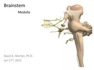
Brainstem • Ventral surface • MEDULLA: • Ventral median fissure: • It divides the medulla into 2 halves. • Its lower part is masked by decussation of pyramidal (corticospinal) fibers. • Pyramid: • It lies on either side of ventral median fissure. • It is an elevation produced by corticospinal tract. Anterior view
Brainstem- Ventral Surface of Medulla • MEDULLA: • Olive: • It lies lateral to the pyramid & separated from it by the ventrolateral sulcus. • It is an elevation produced by inferior olivary nucleus. • Nerves emerging from Medulla (4 nerves): • Hypoglossal (12th): between pyramid & olive • Glossopharyngeal (9th), vagus (10th) & cranial part of accessory (11th): dorsolateral to olive (from above downwards)
Brainstem - Ventral Surface of Pons PONS: • Basilar sulcus • It divides the pons into 2 halves. • It is occupied by basilar artery. • Transverse pontine (pontocerebellar) fibers: • Originate from pontine nuclei. • Cross midline & pass through contralateral middle cerebellar peduncle to enter the opposite cerebellar hemisphere.
Brainstem - Ventral Surface of Pons PONS: • Nerves emerging from Pons (4 nerves): • Trigeminal (5th): from the middle of ventrolateral aspect of pons, as 2 roots: a small medial motor root & a large lateral sensory root • Abducent (6th): at junction between pons & pyramid • Facial (7th) & vestibulocochlear (8th):at cerebellopontine angle (junction between medulla, pons & cerebellum). Both nerves emerge as 2 roots: from medial to lateral:motor root of 7th , sensory root of 7th , vestibular part of 8th & cochlear part of 8th
Brainstem - Ventral Surface of Midbrain • MIDBRAIN: • It is formed of a large column of descending fibers (crus cerebri or basis pedunculi),on either side • The 2 crura cerebri are separated by a depression (interpeduncular fossa) • Nerve emerging from midbrain (one): • Occulomotor (3rd): From medial aspect of crus cerebri
Brainstem - Dorsal Surface of Medulla • Divided into 2 parts: • Caudal 2/3: Closed medulla • Rostral 1/3: Open medulla
Brainstem - Dorsal Surface of Medulla • CLOSED MEDULLA • Containsthe rostral continuation of central canal. • Composed of: • Dorsal median sulcus: divides the closed medulla into 2 halves. • Fasciculus gracilis: on either side of dorsal median sulcus. • Gracile tubercle: an elevation produced at the upper part of fasciculus gracilis, marks the site of gracile nucleus. • Fasciculus cuneatus: on either side of fasciculus gracilis. • Cuneate tubercle:an elevation produced at the upper part of fasciculus cuneatus, marks the site of cuneate nucleus.
Brainstem - Dorsal Surface of Medulla • CLOSED MEDULLA • Composed of: • Obex:(from the Latin for barrier) is the point in the human brain at which the fourth ventricle narrows to become the central canal of the spinal cord. • The obex occurs in the caudal medulla. • Area postrema: The area postrema is situated just before the obex. • The area postrema is a medullary structure in the brain that controls vomiting. • Its privileged location in the brain also allows the area postrema to play a vital role in the control of autonomic functions by the central nervous system.
Brainstem - Dorsal Surface of Medulla • OPEN MEDULLA • Forms the lower part of floor of 4th ventricle. • On either side, an inverted V-shaped sulcus divides the area into 3 parts (from medial to lateral): • Hypoglossal triangle: overlies hypoglossal nucleus • Vagal triangle: overlies dorsal vagal nucleus • Vestibular area: overlies vestibular nuclei
Brainstem - Dorsal Surface of Medulla Lamina tecti (quadrigemina)
Brainstem - Dorsal Surface of Pons • PONS • Forms the upper part of floor of 4th ventricle. • Separated from the medulla by an imaginary line passing between the caudal margins of middle cerebellar peduncle. • On either side, a sulcus divides the area into 2 parts (from medial to lateral): • Medial eminence:overlies abducent nucleus. • Vestibular area: overlies vestibular nuclei.
Brainstem - Dorsal Surface of Pons • Median sulcus: Divides the floor into right and left halves. It extends from cerebral aqueduct of the midbrain to central canal of the spinal cord. • Medial eminence: Elevations on either side of the median sulcus. • Sulcus limitans: medial eminence is laterally bounded by sulcus limitans. • The upper end of the sulcus limitans widens into a triangular depression called "suprior fovea" above suprior fovea sulcus limitans presents a flattend grey area called "locus ceruleus". • The lower end of the sulcus limitans widens into a triangular depression called "Inferior fovea". • Vestibular area:lateral to sulcus limitans vestibular nuclei is overlied by this.
Brainstem - Dorsal Surface of Pons • Locus ceruleus: (also spelled locus coeruleus, or locus caeruleus) • The locus coeruleus is the principal site for brain synthesis of norepinephrine (noradrenaline). • Is a nucleus in the pons involved with physiological responses to stress and panic.
Brainstem - Dorsal Surface of Medulla • Facial colliculus: formed by the internal part of the facial nerve as it loops around the abducens nucleus in the lower pons. • Sulcus limitans: which represents the border between the alar plate and the basal plate of the developing neural tube. • Obex: represents the caudal tip of the fourth ventricle; the obex is also a marker for the level of the foramen magnum of the skull and therefore is a marker for the imaginary dividing line between the medula and spinal cord. • Striamedullaris: fibers derived from arcuate nuclei, which emerge from the median sulcus and run transversely across the floor to enter into the inferior cerebellar penducle.
Brainstem - Dorsal Surface of Midbrain • MIDBRAIN: • Marked by 4 elevations: • Two superior colliculi: concerned with visual reflexes. • Two inferior colliculi: forms part of auditory pathway. • Nerve emerging from midbrain (one): • Trochlear (4th): just caudal to inferior colliculus (the only cranial nerve emerging from dorsal surface of brain stem)
Fourth Ventricle • Cavity of hindbrain • Diamond (rhomboid) in shape • Triangular in cross section • Communications: • Rostrally: with cerebral acqueduct (cavity of midbrain) • Caudally with central canal (cavity of spinal cord) • Lateral walls (boundaries): superior & inferior cerebellar peduncles
Fourth Ventricle • Roof: • Upper part:superior cerebellar peduncle & superior medullary velum (a layer of pia & ependyma bridging the space between the 2 peduncles) • Middle part: cerebellum • Lower part: inferior medullary velum (a layer of pia & ependyma), has a central defect that forms the median aperture of 4th ventricle
Fourth Ventricle • Floor (rhomboid fossa): formed of: • Whole dorsal surface of pons • Open medulla (dorsal surface of rostral 1/3 medulla) • Apertures:provide communication between 4th ventricle & subarachnoid space for circulation of CSF • One median aperture (Foramen of Magendi): in the roof of 4th ventricle • Two lateral apertures (Foramena of Luschka): at cerebellopontine angle
Brainstem - Medulla Cross-section through the medulla at the level of the sensory decussation.
Brainstem - Medulla Cross-section through the medulla at the level of the caudal part of the 4thventricle.
Brainstem - Pons The pons: level of the right VI nerve nucleus and the intrapontine courseof the facial nerve and, on the left, of the nuclei of V.
Brainstem - Midbrain The midbrain: level of the inferior colliculus and decussation of thesuperior cerebellar peduncle.
Brainstem - Midbrain The midbrain: level of the superior colliculus and the red nucleus.
Internal Structures of Brain Stem Gray matter • Cranial nerve nuclei • Relay nuclei
General somatic motor nuclei • Nucleus of oculomotor n. • Nucleus of trochlear n. • Nucleus of abducent n. • Nucleus of hypoglossal n.
Special Visceral Motor Nuclei • Motor nucleus of trigeminal n. • Nucleus of facial n. • Nucleus ambiguus • Accessory nucleus
General Visceral Motor Nuclei • Accessory oculomotor nucleus • Superior salivatory nucleus • Inferior salivertory nucleus • Dorsal nucleus of vagus n.
Visceral Sensory Nuclei (General and special ) • Nucleus of solitary tract
General Somatic Sensory Nuclei • Mesencephalic nucleus of trigeminal n. • Pontine nucleus of trigeminal n. • Spinal nucleus of trigeminal n.
Special Somatic Sensory Nuclei • Cochlear nuclei: SSS- SSA • Vestibular nuclei: SSS- SSA
Relay Nuclei • Gracile nucleus • Cuneate nucleus • Inferior olivary nucleus • Superior olivery nucleus • Pontine nucleus • Nucleus of inferior colliculus • Gray matter layers of superior colliculus • Red nucleus • Substantianigra • Pretectal area
White Matter A lemniscus (Greek for ribbon or band) Ascending tracts • Medial lemniscus • Spinal lemniscus • Trigeminal lemniscus • Lateral lemniscus
White Matter • Medial lemniscus (Reil's band or Reil's ribbon) • The sensory pathway responsible for transmitting: • Fine touch, vibration and conscious proprioceptive information, • As well as tactile pressure, barognosis(the ability of evaluating the weight of objects), graphesthesia(the ability to recognize writing on the skin purely by the sensation of touch), stereognosis(the ability to perceive and recognize the form of an object using cues from texture, size, spatial properties, and temperature), recognition of texture, kinesthesia and two-point discrimination from the body to the cerebral cortex.