Download
1 / 80
800 likes | 1.16k Views
Curriculum Update: SOP and Bradycardia Rhythm Review Based on SOP’s March 2005. Condell Medical Center EMS System October 2006 Site Code #10-7200E-1206 S Hopkins, RN, BSN, EMT-P. Objectives. Upon successful completion of this program, the EMS provider should be able to:

E N D
Curriculum Update:SOP andBradycardia Rhythm ReviewBased on SOP’s March 2005 Condell Medical Center EMS System October 2006 Site Code #10-7200E-1206 S Hopkins, RN, BSN, EMT-P
Objectives • Upon successful completion of this program, the EMS provider should be able to: • identify indications, contraindications, dosing, special considerations, and side effects of medications used in the Region X SOP • participate in rhythm review • state the indications and site of choice of the IO needle • participate in rhythm identification practice • successfully complete the quiz with a score of 80% or greater
Introduction - Adult Intraosseous (IO) Infusion • Can be useful: • when there is a need for IV access and an IV cannot be established in 2 attempts or 90 seconds • May be helpful to use immediately in cardiac arrest or profound hypotension with altered mental status
Adult IO Contraindications • Fracture of tibia or femur (consider alternate extremity) • Infection at intended site • Previous orthopedic procedure to the area (ie: knee replacement, IO previous 480) • Preexisting medical condition (ie: tumor near site, peripheral vascular disease) • Inability to locate landmarks (ie: significant edema) • Excessive tissue at site (ie: morbid obesity)
Adult IO Procedure • BSI protection including face/eye shield • Fill 10 ml syringe with normal saline. • Prime connecting tubing (1 ml) leaving 9 ml in syringe and leave syringe connected to tubing • Identify landmarks • just medial to tibial tuberosity on flat portion of proximal tibia (same site for pediatrics) • FYI: intramedullary vessels do not collapse even in critically ill patients
Adult IO Procedure cont’d • Cleanse insertion site • Prepare EZ-IO driver and needle set • Stabilize leg with non-dominant hand • do not place your hand under patient’s leg • Insert EZ-IO needle at 900 angle
Adult IO Procedure cont’d • Activate driver by depressing trigger on handgrip while maintaining firm & steady pressure on driver • most insertions accomplished under 10 seconds • Once decreased resistance is felt, or needle flange touches skin (whichever is first), release the trigger • While stabilizing hub, remove driver from needle set
Adult IO Procedure cont’d • Remove stylet by rotating counterclockwise • place stylet in sharps container • Connect primed EZ-connect tubing • Using syringe, flush with remaining 10 ml normal saline • observe for swelling or extravasation around site • to improve flow rate, give 10 ml bolus normal saline rapid IVP
Adult IO Procedure cont’d • Confirm needle placement • most reliable indicators: • needle firmly in bone • fluid infuses well • inability to aspirate does not mean non-placement • if placement is in doubt, leave needle in place with connecting tubing & syringe attached and ED staff can reevaluate site
Adult IO Procedure cont’d • Attach EZ-connect to IV tubing & begin infusion • any drug given IVP can be given IO • dosages, onset, & peak concentrations virtually identical to those given IVP • IO route is preferred over ETT route • Apply pressure to IV bag to facilitate flow • flow rates will be slower than IV routes due to anatomy of IO space • pressure may be applied manually or with a blood pressure cuff
Adult IO Procedure cont’d • Secure tubing to leg • Apply wristband supplied with equipment • offers 24 hour hot line for questions • reminds staff to remove EZ-IO within 24 hours • Frequently reassess pressure to IV bag • Monitor EZ-IO site and patient condition • infection rates are low (0.6%) • another EZ-IO may be used in same limb after 48 hours • check calf area for swelling after any fluid bolus
Adult IO Procedure Patient Feedback • “Pain” felt during insertion equivalent to bumping shin on a table (5/10) • lasted < 10 seconds • Similar levels of pain felt when IV infusions started at max rates • your patients will not be conscious! Dr. Miller, EZ-IO developer after practice insertion of device
EZ-IO Device • FYI: • Same drill will eventually be used for pediatric and adult insertion of IO device • Needle size will change to adapt to population receiving IO • Hands-on practice will take place in future CE
Electrical Conduction System • SA • AV node • Bundle of His • Right & Left Bundle Branches • Purkinje Fibers
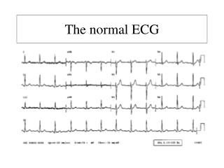
Sinus Bradycardia • Rate: < 60 bpm • Rhythm: regular • P waves: positive, upright, rounded, precede each QRS, all look relatively alike • PR interval: 0.12 - 0.20 seconds; relatively constant • QRS: <0.12 seconds (unless intraventricular conduction delay is present)
Atrioventricular (AV) Blocks • Delay or interruption in impulse conduction in AV node, bundle of His, or His-Purkinje system • Classified according to degree of block and site of block • PR interval is key in determining type of AV block • Width of QRS determines site of block
AV Blocks • Clinical significance dependent on: • degree or severity of the block • rate of the escape pacemaker site • ventricular site will be slower than a junctional site • patient’s response to that ventricular rate • evaluate level of consciousness/responsiveness and blood pressure
Second Degree AV Block Wenckebach, Mobitz Type I • Rate: atrial rate is greater than ventricular rate • Rhythm: atrial rate regular (P to P marches out); ventricular rate irregular (dropped QRS) • P waves: P waves all uniform, not all P waves followed by QRS • PR interval: getting progressively longer until there is a P wave without a QRS • QRS: < 0.12 seconds
Second Degree AV Block Classical, Mobitz Type II • Rate: atrial rate greater than ventricular • Rhythm: atrial regular (P’s to P’s march out); ventricular regular if degree of block is constant • P waves: normal in appearance; not all followed by QRS • PR interval: constant for conducted beats • QRS: < 0.12 seconds
3rd Degree Heart Block - Complete • Rate: atrial rate greater than ventricular; ventricular rate determined by site of escape rhythm • Rhythm: atrial regular (P’s to P’s march out); ventricular regular but no relationship to atrial • P waves: normal in appearance • PR interval: none (no relationship between atrial & ventricular rhythms • QRS: narrow if junctional pacemaker site or wide if ventricular pacemaker site
Helpful Tips • Second degree Type I • think Type I drops one • Wenckebach “winks” when it drops one • Second degree Type II • think 2:1, 2:1, 2:1 • recognize the block can be variable or something other than 2:1 • Third degree - complete • think completely no relationship between atria and ventricles
Junctional Rhythms • Rate: 40 - 60 bpm • Rhythm: very regular • P waves: may occur before, during, or after QRS; if visible are inverted in lead II, III, & AVF • PR interval: if P wave present, usually shortened (< 0.12 seconds) • QRS: normally < 0.12 seconds, longer if aberrantly conducted
Junctional Rhythms Rate determines description: • Junctional escape rhythm rate is 40-60 bpm • Accelerated junctional rhythm rate is 61 - 100 bpm • Junctional tachycardia rate is over 100
Treatment/Interventions Bradycardia • Guided by presence and degree of signs and symptoms • Atropine • used to increase heart rate • can increase rate of SA node discharge; increase speed of conduction through AV node; has little or no effect on contractility • typical dose starts 0.5 mg IVP • maximum dosage 3 mg IVP
Additional Treatment • Transcutaneous pacing • no response to doses of atropine • unstable patient with a wide QRS • set pacing at a rate of 80 beats per minute in the demand mode • start output (mA) at lowest setting possible and increase until capture • Valium 2 mg IVP (increments to 10 mg) should be given to help with the chest discomfort
Patient Unresponsive To Therapy • Consider the patient may be in cardiogenic shock • Consider fluid challenge 200 ml; may repeat once • Evaluate breath sounds before & after fluid • Dopamine drip to maintain B/P >100 • Start dopamine minidrip at 5 mcg/kg/min • Tip - quick drip calculation: take pt’s weight in pounds, take 1st 2 numbers, subtract 2. This is drip factor to start with (ie: pt weight 210#; 21 - 2 = 19; start drip at 19 minidrips/minute)
What Is This Rhythm? • Sinus bradycardia • At this rate the patient is expected to be symptomatic • Treatment if symptomatic? • Atropine for narrow complex QRS; TCP if QRS wide
What Is This Rhythm? • Second degree Type I - Wenckebach • Treatment usually not necessary as heart rate is usually near lower limit of 50’s - 60’s and patient is rarely symptomatic • Monitoring is required for deterioration
What Is This Rhythm? • Second degree Type II - Classical (narrow complex) • Overall ventricular rate is most often slow causing the patient to be symptomatic and requiring therapy
What Is This Rhythm? • Second degree Type II - Classical • Wide QRS indicates the origin of the escape pacemaker site is low down in the conduction system • TCP should be used ASAP if patient symptomatic
What Is This Rhythm? • Third degree heart block - complete • P to P’s are regular; R to R’s are regular • There is no relationship between the atria and ventricles (no pattern or consistency with PR interval)
What Is This Rhythm? • Third degree - complete heart block with a wide QRS complex • Treatment includes avoiding atropine and starting with TCP
What Is This Rhythm? • Junctional rhythm (P waves inverted) • Inherent rate of AV node is 40 -60 bpm • Treatment is based on symptoms and tolerance of patient
What Is This Rhythm? • Second degree Type I - Wenckebach • For some patients, this may be their normal rhythm. For others, they may go back and forth between sinus rhythm and second degree heart block Type I without signs or symptoms
What Is This Rhythm? • Sinus bradycardia with wide QRS (bundle branch block pattern) • Need to determine if patient is symptomatic or not before deciding on interventions needed
What Is This Rhythm? • Third degree heart block - complete • With this appearance and heart rate, patient more than likely will be symptomatic • If narrow QRS, start with atropine • If wide QRS, patient needs TCP (omit atropine)
What Is This Rhythm? • Paced rhythm with single failure to capture • Pacemaker wires may need to be repositioned at the hospital • Carefully monitor EKG for further loss of capture
Revised AHA CPR Guidelines • The message: • focus is “back to basics” • push harder, push faster • 30:2 for adult 1 & 2 man; child & infant 1 man CPR • 15:2 for child & infant 2 man CPR • rate of 100 compressions/minute • perform 5 cycles of 30:2 CPR in 2 minutes and then prepare to defibrillate if needed • switch CPR roles every 2 minutes due to exhaustion (if the compressor is tired, CPR will be sloppy and will not be effective) • minimize CPR interruptions to < 10 seconds
CPR Changes cont’d • perform CPR if there is any delay while charging defibrillator • do not perform pulse checks unless you observe a rhythm that should provide perfusion • after defibrillation immediately resume CPR • do not stop to perform a rhythm check • ventilations over 1 second • once every 5-6 seconds via BVM to mouth • once every 6-8 seconds with advanced airway in place (ETT, combitube, LMA) • IV/IO drug route preferred over ETT route
Review SOG’s • DNR status • properly completed form must be present with patient • can recognize old orange form or new watermelon colored form • can be a reproduction on any color paper • Closest hospital • patient choice when possible & allowable • clinical condition of patient dictating destination • lack of airway • unstable, near arrest condition • psych patient with no preexisting relationship elsewhere
Cardiac Protocol Review • Acute Coronary Syndrome • chew aspirin to enhance absorption • if patient reliable and took daily dose, do not need to repeat dose; inform medical control; if aspirin not given for any reason, document why • if patient < 35, give aspirin and then confer with medical control before giving nitroglycerin or morphine • 12 lead if treating patient for acute coronary syndrome • inform ED you are sending 12 lead
Tachycardia • determine if the patient is stable or unstable • evaluate blood pressure and level of consciousness • if unstable needs cardioversion (start at 100 j) • if stable, determine if QRS is narrow (think adenosine) or wide (think lidocaine) • PEA/asystole • think & treat for potential causes (H’s & T’s) • PEA: epi 1 mg; if rate is <60 atropine 1mg (max 3 mg) • asystole: epi 1 mg; atropine 1 mg (max 3mg)
Stroke/Brain Attack • Screen all patients for time of onset of symptoms • assessment & diagnostics must be completed and drug intervention must be started within 3 hours of onset (>3 hours increases risk of intracranial bleed • Therefore, the most important question is: “What time did your symptoms start?”
Cincinnati Prehospital Stroke Scale • Facial droop • ask patient to smile, big enough to show their teeth • watch for droop and record as right/left sided droop or no droop • Arm drift • ask patient to close their eyes, hold arms out in front, palms up, for 10 seconds • watch for right/left drift or none • Abnormal speech • abnormal is slurring words, using wrong words, or inability to speak
In-Field Spinal Clearance • A reliable patient without signs or symptoms of neck/spine injury and negative mechanism of injury does not require full spinal immobilization • Document findings to support decision to not immobilize • When in doubt, fully immobilize
In-Field Spinal Clearance Criteria • Positive mechanism of injury - immobilize • high velocity MVC >40 mph • unrestrained occupant in MVC • passenger compartment intrusion >12” • ejection from vehicle • rollover MVC • motorcycle collision >20 mph • death in same vehicle • pedestrian struck by vehicle • falls >2 times patient height • diving injury