Download
1 / 1
20 likes | 205 Views
Poly(ADP-ribosylation) in health and disease. Lab: Maria D’Erme & Luciana Mosca. Alessandra Masci PhD Cristina Aureli Italo Tempera PhD . Collaborations : E. Mattia Dept. of Pubblic Health Sciences Unv.”La Sapienza” Roma S. Morano Dept. of Clinical Sciences Unv.”La Sapienza” Roma

E N D
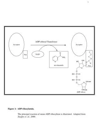
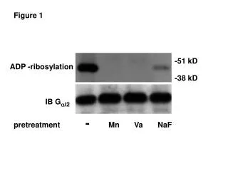
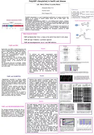
Poly(ADP-ribosylation) in health and disease Lab: Maria D’Erme & Luciana Mosca Alessandra Masci PhD Cristina Aureli Italo Tempera PhD Collaborations: E. Mattia Dept. of Pubblic Health Sciences Unv.”La Sapienza” Roma S. Morano Dept. of Clinical Sciences Unv.”La Sapienza” Roma P. Lieberman Wistar Institute - Philadelphia USA G. Yang Drexel Univ. - Philadelphia USA Poly(ADP-ribosylation) is a post-translational modification of nuclear proteins that appears to be involved in several cellular events such as DNA repair, cell differentiation, apoptosis. The poly(ADP-ribose)polymerase (PARP-1), a zinc-binding nuclear enzyme, catalyzes the covalent addition of the ADP-ribose moiety of nicotinamide adenine dinucleotide to nuclear proteins including histones, transcription factors and PARP itself as well as the subsequent elongation step of the polymer. Futhermore, PARP-1 activation contributes to the development and progression of various pathologies including cancer, diabetes, cerebral ischemia, neurodegenerative diseases and viral infections, highlighting the therapeutic potential of PARP inhibitors in human diseases. Poly(ADP-ribose)polymeration reaction Schematic representation of PARP-1 • Main research fields: • PARP and Epstein Barr Virus: a study on the switch from latent to lytic phase • PARP and type 2 Diabetes: a proteomic approach • PARP and neurodegeneration: use of new PARP inhibitors PARP and EBV Results Epstein Barr virus (EBV), a human herpesvirus wich infects B lymphocytes and pharyngeal ephytelium, is present in roughly 95% of adult individual worlwide. EBV is the ethiological agent of infectious mononucleosis and it is associated with a number of tumors and lymphoproliferative disease in immunocompromized patients. EBV has two distinct cycles of infection: latent, where a limited number of genes is differentially expressed and lytic where all the genes are expressed leading to the production of viral particles. The aim of this work was to investigate whether the poly(ADP-ribosylation) of the cellular insulator CTCF plays a role in the transition from latency to lytic cycle of EBV. To achieve our goals, experiments were performed with both latent and induced Raji cells in the presence or absence of PARP inhibitors. CoIP assay showed that CTCF is polyADP-ribosylated in lytic infected Raji cells, while this isoform was not detected in the viral latent phase. ChIP assay demonstrated that, after the induction of lytic replication, CTCF shows different affinity levels to viral DNA regions than in latent phase.Moreover, immunofluorescence microscopy studies revealed that polyADP-ribosylated CTCF is responsible upon induction, of its traslocation to the nucleolus Figure 1. Lysates obtained from control and induced Raji cells were immunoprecipitated with -CTCF, -PAR and -PARP antibodies. The samples were separated by 10% SDS-PAGE, transferred to PVDF membrane and incubated with respective antibodies. Proteins were visualized by chemiluminescence detection with ECL Advance kit. Figure 2. Real-time PCR analysis of ChIp assay in Mutu1 and Raji cells. After crosslinking with formaldehyde the cells were lysed, sonicated and incubated with a monoclonal anti-CTCF antibody or normal rabbit IgG. The DNA was extracted and analyzed by real-time PCR using a set of primers for Q promoter region of EBV genome. The results are expressed using 2-ct method. PARP and DIABETES Hyperglycemia associated to diabetes mellitus condition increases reactive oxygen species (ROS), Advanced Glycation End-products (AGEs), polyol pathway flux and protein kinase C (PKC) activation, inducing DNA damage at different tissue levels and PARP activation. In different cellular conditions associated with inflammation such as cardiac ischemia, septic and hemorrhagic shock, vascular damage of diabetes, and regeneration of islets of Langerhans, PARP-1 activation has been demonstrated. Since circulating mononuclear cells are involved in inflammation and in the first host response, we intend to analyze, using the proteomic strategies, MNCs from patients with type 2 diabetes Results Figure 3. Immunofluorescence microscopy of CTCF and nucleolin. Control and induced Raji cells were immunostained with primary anti-CTCF antibody (1:50) then with secondary FITC anti-rabbit antibody or with primary anti-nucleolin antibody (1:20) and then with Texas Red-conjugated secondary anti-mouse antibody. The results of immunostaining were observed by fluorescence microscope with AxioCam MRC Zeiss camera (Axiovision 3. 1 software). Western blot analysis of proteomic maps using anti-poly(ADP-ribose) antibodies allows us to reveal a complex pattern of ADP-ribosylated proteins in MNCs and sixteen of the antibodies-reactive spots were identified. Comparison of MNCs poly(ADP-ribosyl)ated proteins between healthy donors and type 2 diabetes patients reveals some differences in the degree of post-translational modification. In particular, the isoforms of GAPDH, of alfa-enolase, of isomerase3P and vimentin exhibit not only a different expression, but also a different level of poly(ADP-ribosyl)ation in diabetic patients compared to control group. Table 1 Protein components of MNCs from NG individuals identified by MALDI-TOF MS Table 2 Protein components of MNCs from DM patients identified by MALDI-TOF MS Fig.1 2D electrophoresis of mononuclear cell lysates of Normoglycemic individuals (NG). Gels were stained with colloidal Coomassie Brilliant Blue G-250. MS-identified proteins are circled and numbered as shown in Tab.1. Fig.2 2D electrophoresis of mononuclear cell lysates of Diabetic patients (DM). Gels were stained with colloidal Coomassie Brilliant Blue G-250. Results The PARP inhibitor capacity of a new 4-chinazolidonic derivative MC2050, has been characterized by evaluating enzyme activity both “in vitro” and in SH-SY5Y neuroblastoma cell line. Its effect has also been assessed in a cellular model which mimicks Alzheimer’s degeneration, i.e. SH-SY5Y cells treated with Amyloid-beta 25-35 fragment (Abeta25-35). The data obtained in vitro by PARP activity colorimetric kit (Trevigen) indicate that the new compound is highly active in the micromolar range (IC50 = 50 microM) compared to other well known inhibitors, chosen as reference drugs. In addition MC2050 is a more potent inhibitor than 3-ABA and PJ34 when assayed in SH-SY5Y cells. Cell viability assays revealed also that this compound is not cytotoxic at the tested concentrations. When the SH-SY5Y cells were pretreated with 10 microM Abeta 25-35, the level of endogenous PARP activity increased up to 40% within 24 hours. In the presence of MC2050 enhancement of PARP activity seems to be blunted. Enolasi Enolasi PARP and NEURODEGENERATION The emerging mechanism dictating the fate of neurons appears to involve the regulation of PAR levels in neurons. Therefore, therapies targeting poly(ADP-ribosyl)ation in the treatment of neurodegenerative conditions such as Alzheimer’s and Parkinson's disease are required to inhibit PAR synthesis and/or facilitate its degradation. GAPDH GAPDH Fig.3 Representative Western blot analysis. Mononuclear cell lysates of NG individuals resolved by 2D SDS-PAGE were transferred to PVDF membrane. The western blots were probed with anti poly(ADP-Ribose) monoclonal antibody. Fig.4 Representative Western blot analysis.Mononuclear cell lysates of DM patients resolved by 2D SDS-PAGE were transferred to PVDF membrane. The western blots were probed with anti poly(ADP-Ribose) monoclonal antibody. Fig1 Cell viability assay. SH-SY5Y neuroblastoma cells were seeded at a density of 15,000 cells/well in 96 wells microplates. After 24 hours the medium was changed to that containing 4 mM 3-ABA, or increasing amounts of MC2050 (25, 50 or 100 uM) and the cells were grown for an additional 24-72 hours. Cell viability was estimated by the MTT method. Fig.2 PARP activity assay. To evaluate whether the Abeta 25-35 amyloid fragment affects PARP activity, enzyme activity was determined by a specifically designed colorimetric kit from Trevigen.