Download
1 / 34
400 likes | 790 Views
An introduction to Endoscopic Ultrasound. Dr Bernard Stacey Consultant Gastroenterologist Southampton. EUS. Equipment Technique Indications Normal anatomy Staging cancers Benign disease. Equipment. Radial Linear. 7.5 and 12 MHz frequencies. Technique. Preparation

E N D
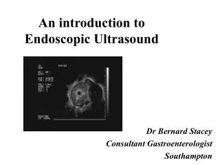
An introduction toEndoscopic Ultrasound Dr Bernard Stacey Consultant Gastroenterologist Southampton
EUS • Equipment • Technique • Indications • Normal anatomy • Staging cancers • Benign disease
Equipment • Radial • Linear
Technique • Preparation • As for normal upper GI endoscopy • Sedation • Alfentanyl and midazolam • Propofol • Antibiotic prophylaxis • Usual indications + biopsy / therapeutics
Indications • Staging cancers • Oesophageal, pancreatico-biliary • Confirming EMR potential • T1 disease, excluding sub-mucosal involvement • Diagnosis and follow up of benign lesions • GIST, lipoma, cysts • Investigating RUQ pain • Investigating pancreatitis
EUS Superficial mucosa / balloon interface Lamina propria Submucosa Balloon Muscularis propria (T2) Adventitia
Staging oesophageal cancer • Part of national guidelines in assessment of potential curable disease • Complementary with CT • T stage • N stage • (M stage)
T4? Abutment echorich plane between tumour and vessel intact Adherence echorich plane partially/totally disrupted >3cm Invasion tumour compressing / growing into vessel lumen
5mm 12mm