Download
1 / 26
780 likes | 3.68k Views
Introduction to Echocardiography Cardiac Ultrasound. Pauline Seydak Clinical Physiology Trainer NI. Echo.

E N D
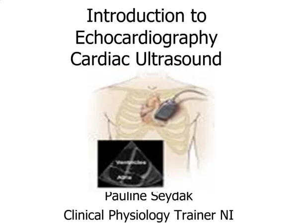
Introduction to EchocardiographyCardiac Ultrasound Pauline Seydak Clinical Physiology Trainer NI
Echo Echo is something you experience all the time. If you shout into a well, the echo comes back a moment later. The echo occurs because some of the sound waves in your shout reflect off a surface (either the water at the bottom of the well or the wall on the far side) and travel back to your ears. A similar principle applies in cardiac ultrasound.
Generation Of An Ultrasound Image Echocardiography (echo or echocardiogram) is a type of ultrasound test that uses high-pitched sound waves to produce an image of the heart. The sound waves are sent through a device called a transducer and are reflected off the various structures of the heart. These echoes are converted into pictures of the heart that can be seen on a video monitor.There is no special preparation for the test.
Cont. Ultrasound gel is applied to the transducer to allow transmission of the sound waves from the transducer to the skin The transducer transforms the echo (mechanical energy) into an electrical signal which is processed and displayed as an image on the screen. The conversion of sound to electrical energy is called the piezoelectric effect
Machines • There are 5 basic components of an ultrasound scanner that are required for generation, display and storage of an ultrasound image. • Pulse generator - applies high amplitude voltage to energize the crystals • Transducer - converts electrical energy to mechanical (ultrasound) energy and vice versa • Receiver - detects and amplifies weak signals • Display - displays ultrasound signals in a variety of modes • Memory - stores video display
Transthoracic Echo A standard echocardiogram is also known as a transthoracic echocardiogram (TTE), or cardiac ultrasound. The subject is asked to lie in the semi recumbent position on his or her left side with the head elevated. The left arm is tucked under the head and the right arm lies along the right side of the body Standard positions on the chest wall are used for placement of the transducer called “echo windows”
Parasternal Long-Axis View (PLAX) Transducer position: left sternal edge; 2nd – 4th intercostal space Marker dot direction: points towards right shoulder Most echo studies begin with this view It sets the stage for subsequent echo views Many structures seen from this view
Parasternal Short Axis View (PSAX) Transducer position: left sternal edge; 2nd – 4th intercostal space Marker dot direction: points towards left shoulder(900 clockwise from PLAX view) By tilting transducer on an axis between the left hip and right shoulder, short axis views are obtained at different levels, from the aorta to the LV apex. Many structures seen
Papillary Muscle (PM)level PSAX at the level of the papillary muscles showing how the respective LV segments are identified, usually for the purposes of describing abnormal LV wall motion LV wall thickness can also be assessed
Apical 4-Chamber View (AP4CH) Transducer position: apex of heart Marker dot direction: points towards left shoulder The AP5CH view is obtained from this view by slight anterior angulation of the transducer towards the chest wall. The LVOT can then be visualised
Apical 2-Chamber View (AP2CH) Transducer position: apex of the heart Marker dot direction: points towards left side of neck (450 anticlockwise from AP4CH view) Good for assessment of LV anterior wall LV inferior wall
Sub–Costal 4 Chamber View(SC4CH) Transducer position: under the xiphisternum Marker dot position: points towards left shoulder The subject lies supine with head slightly low (no pillow). With feet on the bed, the knees are slightly elevated Better images are obtained with the abdomen relaxed and during inspiration Interatrial septum, pericardial effusion, desc abdominal aorta
Suprasternal View Transducer position: suprasternal notch Marker dot direction: points towards left jaw The subject lies supine with the neck hyperexrended. The head is rotated slightly towards the left The position of arms or legs and the phase of respiration have no bearing on this echo window Arch of aorta
Valves of the Heart • Heart valves maintain the unidirectional flow of blood in the heart by opening and closing depending on the difference in pressure on each side. There are four valves in the heart • The two atrioventricular (AV)valves between the atria and the ventricles. • The two semilunar (SL) valves, obvious in the arteries leaving the heart.
Atrioventricular valves(AV) These are small valves that prevent backflow from the ventricles into the atria during systole. They are anchored to the wall of the ventricle by chordae tendineae, that prevent the valve from inverting. The chordae tendineae are attached to papillary muscles that cause tension to better hold the valve. Together, the papillary muscles and the chordae tendinae are known as the subvalvular apparatus. The function of the subvalvular apparatus is to keep the valves from prolapsing into the atria when they close. The subvalvular apparatus have no effect on the opening and closure of the valves. This is caused entirely by the pressure gradient across the valve. AV valves are Mitral and Tricuspid
Mitral Valve (MV) Also known as the bicuspid valve contains two flaps. The mitral valve gets its name from the resemblance to a bishop's mitre(a type of hat). It allows the blood to flow from the left atrium into the left ventricle. It is on the left side of the heart and has two cusps or leaflets, the Anterior MV leaflet (AML) and the Posterior MV leaflet (PMV)
Tricuspid Valve (TV) The tricuspid valve is the three flapped valve on the right side of the heart, between the right atriumand the right ventricle which stops the backflow of blood between the two. This valve consists of 3 leaflets – a large anterior leaflet (ATL), a small septal leaflet (STL) and a tiny posterior leaflet (PTL)
Semilunar Valves These are positioned on the pulmonary artery and the aorta. The semilunar valves are flaps of endocardium and connective tissue reinforced by fibers which prevent the valves from turning inside out. They are shaped like a half moon, hence the name semilunar These valves do not have chordae tendinae. They are named Aortic and Pulmonary
Aortic Valve (AV) Lies between the left ventricle and the aorta and has three cusps, anterior right coronary cusp (RCC) posterior non-coronary cusp (NCC) middle left coronary cusp (LCC) During ventricular systole, pressure rises in the left ventricle. When the pressure in the left ventricle rises above the pressure in the aorta, the aortic valve opens, allowing bloodto exit the left ventricle into the aorta.
Pulmonary Valve (PV) Lies between the right ventricle and the pulmonary artery and has three cusps a posterior (left) cusp, an anterior cusp and a right cusp. Similar to the aortic valve, the pulmonary valve opens in ventricular systole, when the pressure in the right ventricle rises above the pressure in the pulmonary artery. At the end of ventricular systole, when the pressure in the right ventricle falls rapidly, the pressure in the pulmonary artery will close the pulmonary valve.
The Modalities of Echo • The following modalities of echo are used clinically: • Conventional echo • Two-Dimensional echo (2-D echo) • Motion- mode echo (M-mode echo) • Doppler Echo • Continuous wave (CW) Doppler • Pulsed wave (PW) Doppler • Colour flow(CF) Doppler • All modalities follow the same principle of ultrasound • Differ in how reflected sound waves are collected and analysed
Two-Dimensional Echo (2-D echo) This technique is used to "see" the actual structures and motion of the heart structures at work. Ultrasound is transmitted along several scan lines(90-120), over a wide arc(about 900) and many times per second. The combination of reflected ultrasound signals builds up an image on the display screen. A 2-D echo view appears cone-shaped on the monitor.
M-Mode echocardiography An M- mode echocardiogram is not a "picture" of the heart, but rather a diagram that shows how the positions of its structures change during the course of the cardiac cycle. M-mode recordings permit measurement of cardiac dimensions and motion patterns. Also facilitate analysis of time relationships with other physiological variables such as ECG, and heart sounds.
Doppler echocardiography Doppler echocardiography is a method for detecting the direction and velocity of moving blood within the heart. Pulsed Wave (PW) useful for low velocity flow e.g. MV flow Continuous Wave (CW) useful for high velocity flow e.g aortic stenosis Color Flow (CF) Different colors are used to designate the direction of blood flow. red is flow toward, and blue is flow away from the transducer with turbulent flow shown as a mosaic pattern.
Conclusion Echocardiography provides a substantial amount of structural and functional information about the heart. Still frames provide anatomical detail. Dynamic images tell us about physiological function The quality of an echo is highly operator dependent and proportional to experience and skill, therefore the value of information derived depends heavily upon who has performed it