Download
1 / 54
870 likes | 1.61k Views
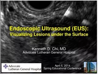
Endoscopic Ultrasound (EUS): Visualizing Lesions under the Surface. Kenneth D. Chi, MD Advocate Lutheran General Hospital. April 5, 2014 Spring Educational Conference. Outline. 1. Basic primer in EUS How has EUS changed patient care and community referrals?

E N D
Endoscopic Ultrasound (EUS): Visualizing Lesions under the Surface Kenneth D. Chi, MDAdvocate Lutheran General Hospital April 5, 2014Spring Educational Conference
Outline 1. Basic primer in EUS • How has EUS changed patient care and community referrals? • When do you refer for an EUS? What is appropriate referral? 4. When is EUS useful? / What are limitations / Complications? 5. Applications of EUS at Lutheran General Hospital 6. Future Applications of EUS
What is EUS? • Endoscopic Ultrasound has expanded the breadth of GI Endoscopy • Introduced in 1980s: Japan / USA / Germany • Able to visualize pancreas through the stomach wall • Permits detailed imaging of GI wall layers • Enables accurate locoregional tumor staging
The EUS Scopes Miniprobe Linear (FNA) Radial
Radial vs. Linear Yusuf, et al. Gastrointest Endosc. 2007 Jul;66(1):131-43.
Basic principles of Ultrasound Hyper-echoic (bright) Hypo-echoic (dark) An-echoic (black) Iso-echoic (same) Yusuf, et al. Gastrointest Endosc. 2007 Jul;66(1):131-43.
(mucosa) (muscularis mucosa) (submucosa) (muscularis propria) (adventitia / serosa)
How EUS has changed patient care Esophageal cancer staging: EUS results could dramatically change the patient’s treatment course ? ?
Role of EUS in Esophageal Ca • Central role in initial staging as outcome is strongly associated with stage • Useful in monitoring disease recurrence • Has complementary role with other imaging: • EUS for locoregional staging • CT / PET : eval for mets / stage IV dz
Comparing CT scan vs. EUS in detecting Lymph Nodes ( Lymph node staging in Esophageal Cancer) Vazquez-Sequeiros, E, Clain, JE, Norton, ID, et al, Gastroenterology 2003; 125:1626.
Esophageal Cancer Staging Algorithm Primary Diagnosis (EGD) Resectable Disease CT Scan (+/- PET) EUS Unresectable Disease T4 or M1 Stage Dependent Treatment T1 (T2) N0 T3 or TxN1 T4 or M1 ChemoXRT Palliation Surgical Resection Chemo / XRT Resection
EUS T + N Staging T1 T4 T2 T3 EUSLayer 5 4 3 2 1
Why is T Stage Important?Risk of LN Mets Depth of tumor predicts LN involvement Compared to T1 patient: T2 = 6x more likely to have N1 T3 = 23x T4 = 35x Rice, TW et. al Ann Thorac Surg. 1998 Mar;65(3):787-92.
Clinical impact of EUS EUS FNA *In this study, EUS/FNA dramatically changed 20% (5/7) patients management course Shami VM, Villaverde A, Stearns L, Chi KD, Kinney TP, Rogers GB, Dye CE, Waxman I. Endoscopy. 2006 Feb;38(2):157-61.
Cost analysis of EUS Impact of pre-op EUS on Esophageal cancer management and cost • 26% of patients undergoing pre-op EUS staging would be spared combined modality therapy who were found to be Stage I or IV. In other words: • Estimated for every 100 pts undergoing pre-op EUS for Esophageal cancer staging: • 14 pts with Stage I would be spared neo-adjuvant CTX (Total Cost savings $122,192) • 12 pts with Stage IV would be spared surgery (saving a total of $285,600) • Average cost savings $3443 per patient (Shumaker, et. al Gastrointest Endosc. 2002 Sep;56(3):391-6.)
EUS Indications Question: Are community physicians aware of the indications of EUS?
EUS Indications / Limitations • 1st study to assess knowledge of referring indications of EUS among physicians • Setting: Mayo Clinic, Rochester • 25 question survey • Surveyed: 121 GI 259 Internists 129 non-GI subspecialties 150 Surgeons Yusuf TE et. al, GIE 2004;60:575-9.
Average Score per Specialty Yusuf TE et. al, Gastrointest Endosc 2004;60:575-9.
What does this mean? • Gastroenterologists still responded incorrectly to 15% of questions • Liver, Pancreas, and Lower intestine EUS were the least understood among referrers • More education is needed regarding EUS use and it’s limitations
Use of EUS at LGH Utilization of EUS for locoregional staging for Esophageal Cancer & GEJ CA • LGH Data 2005-2007. EUS Available at LGH 1/2005.
Limitations of EUS • Ultrasound can only “see so far” • Time-consuming. • Doing EUS when there is no target lesion is like looking for a needle in a haystack. • Technical challenges: • Altered anatomy • Small mucosal lesions • Non-diagnostic FNA passes • Newer FNA needles allowing “core biopsies” for pathology • On-site cytopatholgist improves diagnostic yield of EUS-FNA • (Klapman JB et al., Am J Gastroenterol. 2003 Jun;98(6):1289-94. )
Complications of EUS • Infection risk after FNA • Primarily in pancreatic cyst aspiration • Studies show bacteremia incidence of 0.4% - 1% (Voss et al. Gut 2000:46:244-9) • IV antibiotic pre/post procedure • Bleeding • Mild intraluminal bleeding: 4% (Voss et al. Gut 2000:46:244-9) • Extraluminal bleeding: 1.3% (Affi et al. GIE 2001; 53:221-5) • Perforation • Standard EGD risk: 0.03% (Eisen et al. GIE 2002; 55:784-93) • Diagnostic EUS risk: 0.07% (Rahod & Maydeo GIE 2002; 56:AB169) • Pancreatitis after EUS/FNA: 1%-2%(Gress et al. GIE 2002;56:864-7) • EUS is very safe; Similar risks to diagnostic EGD
Applications of EUS at LGH • Esophageal cancer locoregional staging • “Abnormal CT scan” – pancreatic lesion • Solid & cystic pancreatic lesions • Pancreatic cyst fluid analysis • Mediastinallymphadenopathy(with EBUS) • Evaluation of submucosal lesions • Difficult polypectomy cases • Evaluation prior to EMR • Celiac plexus neurolysis • EUS-guided Pancreatic pseudocyst drainage • EUS-guided “Rendez-vous” ERCP • Rectal EUS
EUS guided Celiac Plexus Neurolysis • Pancreatic cancer: • Pain score reduction in 78% of pts at 2 wks, and sustained for 24 wks • Chronic Pancreatitis: • Pain score reduction in 50% of pts and sustained for 24 wks.
Utilizing EUS in Polypectomy • 43 y.o. athlete referred to evaluate incidental antral nodule found on EGD during workup of abdominal pain.
Utilizing EUS in Polypectomy Marking Borders Saline Lift
Utilizing EUS in Polypectomy Snare within Cap Resection Site
Localization of Neuroendocrine Tumor 2006 - EGD
Localization of Neuroendocrine Tumor 2008 - EGD
Localization of Neuroendocrine Tumor 2008 - EGD
Localization of Neuroendocrine Tumor 5/29/2008 - EUS
Localization of Neuroendocrine Tumor 5/29/2008 - EUS FNA revealed neuroendocrine cells consistent with Gastrinoma
EUS-guided cystgastrostomy in Pancreatic pseudocyst drainage
EUS-guided Rendezvous • 47 y.o. woman with symptomatic pancreas divisum for minor papilla
EUS-guided Rendezvous Failed ERCP attempt of minor papilla
EUS-guided Rendezvous Dilated main pancreatic duct
EUS-guided Rendezvous Transgastric access of main pancreatic duct
EUS-guided Rendezvous Trans-gastric puncture into PD
EUS-guided Rendezvous Trans-gastric puncture into PD
EUS-guided Rendezvous Guidewire puncture into stomach Wire exiting minor papilla