Download
1 / 62
620 likes | 642 Views
Explore the complex mechanisms of neural development from embryo stage to fine-tuning, uncovering how the brain wires and processes sensory information. Discover the principles of neural science and the role of activity-dependent tuning in shaping brain functionality and connectivity. ####

E N D
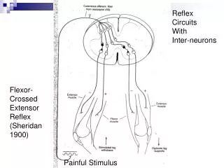
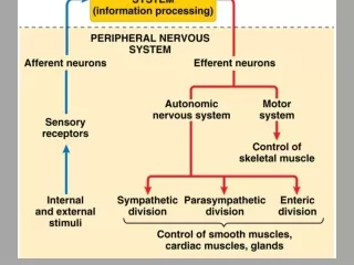
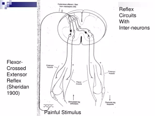
Reflex Circuits With Inter-neurons Flexor- Crossed Extensor Reflex (Sheridan 1900) Painful Stimulus
A Simple model or Minimum Circuit: HIGHER LEVEL CONTROL Pain Stimulus MotoNeuron + + + Sensory Neuron Inter Neuron Inter Neuron Flexor MotoNeuron - - Inter Neuron Inter Neuron Extensor
Neural Development CS182/CogSci110/Ling109 Spring 2007 Jerry Feldman feldman@icsi.berkeley.edu
Lecture Overview • Summary Overview • Development from embryo • Initial wiring • Activity dependent fine tuning • Additional information/details • Principles of Neural Science. Kandel, Schwartz, Jessell, Mcgraw-Hill (2000).
How does this happen • Many mechanisms of human brain development remain unclear, but • Neuroscientists are beginning to uncover some of these complex steps through studies of • the roundworm, fruit fly, frog, zebrafish, mouse, rat, chicken, cat and monkey. • Many initial steps in brain development are similar across species, while later steps are different. • By studying these similarities and differences, we can learn how the human brain develops and hopefully how brain abnormalities, such as mental retardation and other brain disorders, can be prevented or treated.
Summary Overview • The Amoeba (ref. book) uses • complex sensing molecules penetrating its cell membrane to trigger • chemical mechanisms that cause it to move its blobby body towards food and away from harmful substances. • Neurons are also cells and, • in early development, behave somewhat like Amoeba in approaching and avoiding various chemicals. • But rather than the whole cell moving, neural growth involves the outreach of the cell’s connecting pathway (axon) towards its downstream partner neurons.
Summary Overview • The basic layout of visual and other maps is established during development by millions of neurons • each separately following a pattern of chemical markers to • its pre-destined brain region and specific sub-areas within that region. • For example, a retinal cell that responds best to red light • in the upper left of the visual field will connect to cells in the brain that are tuned to the same properties and • these cells, in turn, will link to other cells that use these particular properties giving rise to specific connectivity and cell receptive field properties (topographic maps).
Summary Overview • In the course of development, • detector molecules in the growing neuron interact with • guide molecules to route the connection to the right general destination, sometimes over long distances as in the connection from the spinal cord to the knee. • This process will get neural connections to the right general area, but • aligning the millions of neurons in visual and other neural maps also involves • chemical gradients, again utilizing mechanisms that are very old in evolutionary terms.
Summary Overview • When an axon tip gets to an appropriately marked destination cell, the contact starts a process that develops rudimentary synapses. • Local competition among neural axons with similar marker profiles produces some further tuning at the destination.
Summary Overview:Activity Dependent Tuning • In fact, the initial wiring is only approximate and leaves each neuronal axon connected to several places in the neighborhood of each of its eventual partner neurons. • A second, activity dependent, mechanism is required to complete the development process. • The initial chemical wiring actually produces many more connections and somewhat more neurons than are present in adult brains. • The detailed tuning of neural connections is done by eliminating the extra links, as well as the strengthening functional synapses based on neural activity.
Summary • Neural development and learning is moving from a mystery to routine science. • We know enough to shape theories of how our brains learn skills, including language, and how we acquire and use knowledge. • Contemporary theories of learning are also heavily influenced by psychological experiments, some of which are described next week.
Lecture Overview • Summary Overview • Development from embryo • Neural tube development • Cell division and neuronal identity • Mechanisms for cell type formation and communication • Initial wiring • Activity dependent fine tuning • Plasticity and Learning
Development from Embryo • The embryo has three primary layers that undergo many interactions in order to evolve into organ, bone, muscle, skin or neural tissue. • The outside layer is the ectoderm • (skin, neural tissue), • the middle layer is the mesoderm • (skeleton, cardiac) and • inner layer is the endoderm • (digestion, respiratory).
Neural Tissue • The skin and neural tissue arise from a single layer, known as the ectoderm • in response to signals provided by an adjacent layer, known as the mesoderm. • A number of molecules interact to determine whether the ectoderm becomes neural tissue or develops in another way to become skin
In humans, during the 3rd week, this mesoderm begins to segment. The neural plate folds to form a neural groove and folds.
Other structures including heart, Skeleton, etc. The neural groove fuses dorsally to form a tube at the level of the 4th somite and "zips up” cranially and caudally and the neural crest migrates into the mesoderm (somites differentiate to form vertebrae, muscles).
BRAIN DEVELOPMENT. The human brain and nervous system begin to develop at three weeks’ gestation as the closing neural tube (left). By four weeks, major regions of the human brain can be recognized in primitive form, including the forebrain, midbrain, hindbrain, and optic vesicle (from which the eye develops). Irregular ridges, or convolutions, are clearly seen by six months.
Lecture Overview • Summary Overview • Development from embryo • Neural tube development • Cell division and neuronal identity • Cell communication • Initial wiring • Activity dependent fine tuning • Plasticity and Learning • Development and Infant behavior
Neural cell categories • After the ectodermal tissue has acquired its neural fate, • another series of signaling interactions determine the type of neural cell to which it gives rise. • The mature nervous system contains a vast array of cell types, which can be divided into two main categories: • the neurons, primarily responsible for signaling, • and supporting cells called glial cells.
Factors/gradients in cell formation • Researchers are finding that the destiny of neural tissue depends on a number of factors, including position, that define the environmental signals to which the cells are exposed. • For example, a key factor in spinal cord development is a secreted protein called sonic hedgehog that is similar to a signaling protein found in flies. • The protein, initially secreted from mesodermal tissue lying beneath the developing spinal cord, marks young neural cells that are directly adjacent to become a specialized class of glial cells. • Cells further away are exposed to lower concentrations of sonic hedgehog protein, and they become the motor neurons that control muscles. • An even lower concentration promotes the formation of interneurons that relay messages to other neurons, not muscles.
Timing of Cell Differentiation • Remarkably, the final position of the neuron (its laminar position) is correlated exactly to its birthdate • The birthdate is the time of final mitosis • Cells leaving later migrate past the older neurons (in deeper cortical layers) to the outermost cortex. • The layering of the cortex is thus an inside-first outside-last layering.
Lecture Overview • Summary Overview • Development from embryo • Initial Wiring details • Axon Guidance • Synapse formation • Activity dependent fine tuning • Plasticity and Learning • Development and Infant behavior
Axon guidance mechanisms • Axonal growth is led by growth cones • Filopodia (growing from axons) are able to sense the environment ahead for chemical markers and cues. • Mechanisms are fairly old in evolutionary terms. • Intermediate chemical markers • Guideposts studied in invertebrates • Short and long range cues • Short range chemo-attraction and chemo-repulsion • Long range chemo-attraction and chemo-repulsion • Gradient effects
Axons locate their target tissues by using chemical attractants (blue) and repellants (orange) located around or on the surface of guide cells. Left: An axon begins to grow toward target tissue. Guide cells 1 and 3 secrete attractants that cause the axon to grow toward them, while guide cell 2 secretes a repellant. Surfaces of guide cells and target tissues also display attractant molecules (blue) and repellant molecules (orange). Right: A day later, the axon has grown around only guide cells 1 and 3.
Synapse formation • The two cells exchange a variety of signals. • Vesicles cluster at the pre-synaptic site • Transmitter receptors cluster at the post-synaptic site. • The Synaptic Cleft forms • When the growth cone contacts the target cell (immature muscle cell in the case of a motor neuron), a cleft (basal lamina) forms. • Multiple growth cones (axons) get attracted to the cleft. • All but one axon is eliminated. • A myelin sheath forms around the synaptic cleft and the synaptic connection is made.
Basic Process • Neurons are initially produced along the central canal in the neural tube. • These neurons then migrate from their birthplace to a final destination in the brain. • They collect together to form each of the various brain structures and acquire specific ways of transmitting nerve messages. • Their processes, or axons, grow long distances to find and connect with appropriate partners, forming elaborate and specific circuits. • Finally, sculpting action eliminates redundant or improper connections, honing the specificity of the circuits that remain. • The result is the creation of a precisely elaborated child’s network of 100 billion neurons capable of body movement, perception, an emotion or a thought.
Nature requires Nurture • Initial wiring is genetically controlled • Sperry Experiment • But environmental input critical in early development • Occular dominance columns • Hubel and Wiesel experiment
Sperry’s experiment • Each location in space is seen by a different location on the retina of the frog • Each different location on the retina is connected by the optic nerve to a different location in the brain • Each of these different locations in the brain causes a different movement direction. • In a normal animal, a retinal region which sees in a particular direction is connected to a tectal region which causes a movement in that direction
Innervation of the Optic tectum • Ganglion Cells in the retina map systematically to cells in the optic tectum. • The image of the external stimulus is inverted in the retina and the mapping from the retina to the optic tectum reverts to the original image. • The Nasal ganglion cells of the retina map to the posterior region of the Optic tectum and the temporal ganglion cells map to the anterior region of the tectum
Sperry’s experiment • Sperry took advantage of the fact that in amphibians, the optic nerve will regrow after it has been interrupted • Sperry cut the optic nerve and simultaneously rotated the eye 180 degrees in the eye socket. • In 'learning’ movements to catch prey, the part of the retina now looking forward (backward) should connect to the part of the brain which causes forward (backward) movement.
Sperry’s findings • After regeneration, • his animals responded to prey items in front by turning around and • to prey items behind by moving forward. and • kept doing this even though they never succeeded in reaching the prey.
Conclusion from experiment • The conclusion from this (and some supporting experiments) is • that the pattern of connections between retina and tectum, and the movement information represented is not based on experience. • It is innate based on the initial distribution of chemical markers in the brain.
Lecture Overview • Summary Overview • Development from embryo • Initial wiring • Activity dependent fine tuning
The role of the environment • The development of ocular dominance columns • Cat and later monkey (Hubel and Wiesel) • Retinal input connects to the LGN (Thalamus) • LGN is composed of layers. Each layer receives input (axons) from a single eye • LGN connects to layer IV of the visual cortex • The visual cortex develops ocular dominance columns • Cells that are connected to similar layers in the LGN get stacked together in columns forming stripes. http://neuro.med.harvard.edu/site/dh/
LGN VISUAL CORTEX
Monocular deprivation critical period • Hubel and Wiesel deprived one of the eyes of the cat (later macaque monkey) at various times 1 week – 12 weeks (in the monkey case) 4 weeks – 4 months (for the cat). • The found that the ocular dominance cell formation was most severely degraded if deprivation occurred at 1 – 9 weeks after birth. • Deprivation after the plastic period had no long-term effect.