Download
1 / 1
20 likes | 117 Views
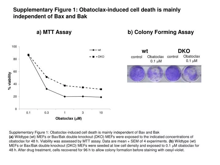
Supplementary Figure 1: Obatoclax-induced cell death is mainly independent of Bax and Bak. a) MTT Assay. b) Colony Forming Assay. wt. DKO. Obatoclax 0.1 m M. control. Obatoclax 0.1 m M. control. Supplementary Figure 1: Obatoclax-induced cell death is mainly independent of Bax and Bak

E N D
Supplementary Figure 1: Obatoclax-induced cell death is mainly independent of Bax and Bak a) MTT Assay b) Colony Forming Assay wt DKO Obatoclax 0.1 mM control Obatoclax 0.1 mM control Supplementary Figure 1: Obatoclax-induced cell death is mainly independent of Bax and Bak (a) Wildtype (wt) MEFs or Bax/Bak double knockout (DKO) MEFs were exposed to the indicated concentrations of obatoclax for 48 h. Viability was assessed by MTT assay. Data are mean + SEM of 4 experiments. (b) Wildtype (wt) MEFs or Bax/Bak double knockout (DKO) MEFs were seeded at low cell density and exposed to 0.1 mM obatoclax for 48 h. After drug treatment, cells recovered for 96 h to allow colony formation before staining with cesyl-violet.