Download
1 / 63
640 likes | 809 Views
Enzyme Specificity and Regulation Reginald Garrett and Charles Grisham. Outline. 15.1 Specificity from Molecular Recognition 15.2 Controls over Enzymatic Activity 15.3 Allosteric Regulation of Enzyme Activity 15.4 Allosteric Model 15.5 Glycogen Phosphorylase

E N D
Enzyme Specificity and Regulation Reginald Garrett and Charles Grisham
Outline • 15.1 Specificity from Molecular Recognition • 15.2 Controls over Enzymatic Activity • 15.3 Allosteric Regulation of Enzyme Activity • 15.4 Allosteric Model • 15.5 Glycogen Phosphorylase • SPECIAL FOCUS: Hemoglobin and Myoglobin
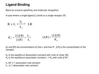
15.1 Specificity The Result of Molecular Recognition • Substrate (small) binds to enzyme (large) via weak forces - what are they? • H-bonds, van der Waals, ionic • sometimes hydrophobic interactions • Understand the lock-and-key and induced-fit models • Relate induced-fit to transition states
15.2 Controls over Enzyme Activity Six points: • Rate slows as product accumulates • Rate depends on substrate availability • Genetic controls - induction and repression • Enzymes can be modified covalently • Allosteric effectors may be important • Zymogens, isozymes and modulator proteins may play a role
15.3 Allosteric Regulation Action at "another site" • Enzymes situated at key steps in metabolic pathways are modulated by allosteric effectors • These effectors are usually produced elsewhere in the pathway • Effectors may be feed-forward activators or feedback inhibitors • Kinetics are sigmoid ("S-shaped")
Models for Allosteric Behavior • Monod, Wyman, Changeux (MWC) Model: allosteric proteins can exist in two states: R (relaxed) and T (taut) • In this model, all the subunits of an oligomer must be in the same state • T state predominates in the absence of substrate S • S binds much tighter to R than to T
More about MWC • Cooperativity is achieved because S binding increases the population of R, which increases the sites available to S • Ligands such as S are positive homotropic effectors • Molecules that influence the binding of something other than themselves are heterotropic effectors
Glycogen PhosphorylaseAllosteric Regulation and Covalent Modification • GP cleaves glucose units from nonreducing ends of glycogen • A phosphorolysis reaction • Muscle GP is a dimer of identical subunits, each with PLP covalently linked • There is an allosteric effector site at the subunit interface
Glycogen PhosphorylaseAllosteric Regulation and Covalent Modification • Pi is a positive homotropic effector • ATP is a feedback inhibitor, and a negative heterotropic effector • Glucose-6-P is a negative heterotropic effector (i.e., an inhibitor) • AMP is a positive heterotrophic effector (i.e., an activator)
Regulation of GP by Covalent Modification • In 1956, Edwin Krebs and Edmond Fischer showed that a ‘converting enzyme’ could convert phosphorylase b to phosphorylase a • Three years later, Krebs and Fischer show that this conversion involves covalent phosphorylation • This phosphorylation is mediated by an enzyme cascade (Figure 15.19)
cAMP is a Second Messenger • Cyclic AMP is the intracellular agent of extracellular hormones - thus a ‘second messenger’ • Hormone binding stimulates a GTP-binding protein (G protein), releasing G(GTP) • Binding of G(GTP) stimulates adenylyl cyclase to make cAMP
Hemoglobin A classic example of allostery • Hemoglobin and myoglobin are oxygen transport and storage proteins • Compare the oxygen binding curves for hemoglobin and myoglobin • Myoglobin is monomeric; hemoglobin is tetrameric • Mb: 153 aa, 17,200 MW • Hb: two alphas of 141 residues, 2 betas of 146
Hemoglobin FunctionHb must bind oxygen in lungs and release it in capillaries • When a first oxygen binds to Fe in heme of Hb, the heme Fe is drawn into the plane of the porphyrin ring • This initiates a series of conformational changes that are transmitted to adjacent subunits
Hemoglobin FunctionHb must bind oxygen in lungs and release it in capillaries • Adjacent subunits' affinity for oxygen increases • This is called positive cooperativity
Myoglobin Structure Mb is a monomeric heme protein • Mb polypeptide "cradles" the heme group • Fe in Mb is Fe2+ - ferrous iron - the form that binds oxygen • Oxidation of Fe yields 3+ charge - ferriciron -metmyoglobin does not bind oxygen • Oxygen binds as the sixth ligand to Fe • See Figure 15.26 and discussion of CO binding
The Conformation Change The secret of Mb and Hb! • Oxygen binding changes the Mb conformation • Without oxygen bound, Fe is out of heme plane • Oxygen binding pulls the Fe into the heme plane • Fe pulls its His F8 ligand along with it • The F helix moves when oxygen binds • Total movement of Fe is 0.029 nm - 0.29 A • This change means little to Mb, but lots to Hb!
Binding of Oxygen by Hb The Physiological Significance • Hb must be able to bind oxygen in the lungs • Hb must be able to release oxygen in capillaries • If Hb behaved like Mb, very little oxygen would be released in capillaries - see Figure 15.22! • The sigmoid, cooperative oxygen binding curve of Hb makes this possible!
Oxygen Binding by Hb A Quaternary Structure Change • When deoxy-Hb crystals are exposed to oxygen, they shatter! Evidence of a structural change! • One alpha-beta pair moves relative to the other by 15 degrees upon oxygen binding • This massive change is induced by movement of Fe by 0.039 nm when oxygen binds • See Figure 15.32